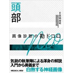
頭部 画像診断の勘ドコロNEO

出版社: メジカルビュー社
著者:
発行日: 2021-02-20
分野: 臨床医学:一般  >  画像/超音波
ISBN: 9784758316132
シリーズ: 画像診断の勘ドコロ
電子書籍版: 2021-02-20 (第1版第2刷)
書籍・雑誌
≪全国送料無料でお届け≫
取寄せ目安:約3営業日

9,680 円(税込)

電子書籍
章別単位での購入はできません
ブラウザ、アプリ閲覧

9,680 円(税込)

商品紹介

「画像診断の勘ドコロNEO」シリーズ第2弾は頭部。
「なぜそうみえるのか」がわかるモダリティの最重要ポイントに加え,スペシャリストたちが読影室で「どこを見て」「どう鑑別し」「どう診断しているのか」,多数の写真と明快な文章で解説する。項目別の「ここが勘ドコロ」など,スペシャリストたちからのアドバイスも満載し,頭部の勉強にも読影室で困ったときにも使える充実の一冊。
発展を続ける画像技術の要点やCOVID-19脳症を含めた最新の知見も網羅。執筆陣渾身の実践知が冴える,白熱の神経放射線読影教室。

目次

  • 第1章 基礎編
     01 正常解剖

    第2章 モダリティ編
     01 CT 
     02 MRI通常画像/読影に関する総論
     03 MRA
     04 拡散MR画像
     05 灌流
     06 磁化率・位相画像
     07 造影剤
     08 定量・合成MRI:Synthetic MRI/MR Fingerprinting
     09 MRS
     10 核医学

    第3章 疾患編
     01 脳血管障害(虚血性疾患)
     02 脳血管障害(出血性疾患)
     03 免疫系の異常に関連する疾患
     04 中毒/治療関連/外因性代謝疾患
     05 遺伝子障害等,内因性の代謝異常
     06 変性疾患・蓄積病
     07 感染症・感染に伴う脳症
     08 脳腫瘍(グリオーマなど)
     09 脳腫瘍(グリオーマ以外)
     10 下垂体・傍鞍部
     11 小児脳の発達と特徴/周産期障害
     12 脳の先天奇形と神経皮膚症候群
     13 てんかん
     14 脳脊髄液腔の異常
     15 外傷

この書籍の参考文献

参考文献のリンクは、リンク先の都合等により正しく表示されない場合がありますので、あらかじめご了承下さい。

本参考文献は電子書籍掲載内容を元にしております。

第1章 基礎編

P.13 掲載の参考文献
牧 豊ほか:神経放射線学(I)(II), 朝倉書店, 東京, 1979.
御供政紀:頭部CT徹底診断, 医学書院, 東京, 1992.
P.14 掲載の参考文献
Otake S, Taoka T, Maeda M, Yuh, W:A guide to identification and selection of axial planes in magnetic resonance imaging of the brain. Neuroradiol J, 31:336-44, 2018.

第2章 モダリティ編

P.16 掲載の参考文献
1. 山口 功ほか:CT撮影技術学 改訂3版, オーム社, 東京, 2017, p18-29.
P.17 掲載の参考文献
2. 公益社団法人日本放射線技術学会:X線CT撮影技術における標準化-GALACTIC 改訂2版, 日放線技会誌, 8, 137-203, 2016.
P.18 掲載の参考文献
3. Boone JM, et al:An accurate method for computer-generating tungsten anode x-ray spectra from 30 to 140kV. Medical Physics, 24:1661-1670, 1997.
P.19 掲載の参考文献
4. 村松禎久ほか:学術調査研究班報告 CT用自動露出機構(CT-AEC)の性能評価班報告書. 日放線技会誌, 61:1094-1103, 2005.
5. 村松禎久ほか:学術調査研究班報告 CT用自動露出機構(CT-AEC)の性能評価班 最終報告書. 日放線技会誌, 63:534-545, 2007.
6. Berrington de Gonzalez A, et al:Risk of cancer from diagnostic X-rays:estimates for the UK and 14 other countries. Lancet, 363:345-351, 2004.
7. ICRP Publication 87:Managing patient dose in multi-detector computed tomography. Ann ICRP, 30, 2000.
P.20 掲載の参考文献
8. AAPM Report No. 204:Size-Specific Dose Estimates(SSDE)in Pediatric and Adult Body CT Examinations,:2011.
9. AAPM Report No. 220:Use of Water Equivalent Diameter for Calculation Patient Size and Size-Specific Dose Estimates(SSDE)in CT, 2014.
P.22 掲載の参考文献
10. 金森勇雄:第4章マルチスライスCT装置. 改訂X線CTの実践, 医療科学社, 東京, 51-58, 20. www.iryokagaku.co.jp/frame/03-honwosagasu.
P.23 掲載の参考文献
11. 浜口直子ほか:急性期脳梗塞診断に対するヘリカルスキャンの適用に関する検討. 日放線技会誌, 66:632-640, 2010.
P.24 掲載の参考文献
12. Lev MH et al:Acute stroke:improved nonenhanced CT detection:benefits of soft-copy interpretation by using variable window width and center level settings). Radiology, 213:150-155, 1999.
P.26 掲載の参考文献
13. Watanabe Y. et al:Dual-energy direct bone removal CT angiography for evaluation of intracranial aneurysm or stenosis:comparison with conventional digital subtraction angiography. Eur Radiol, 19:1019-1024, 2009.
Gupta R, et al:Evaluation of dual-energy CT for differentiating intracerebral hemorrhage from iodinated contrast material staining. Radiology, 257:205-211, 2010.
P.29 掲載の参考文献
14. Smits M:Imaging of oligodendroglioma. Br J Radiol. 89:20150857, 2016.
P.31 掲載の参考文献
14. Yuan MK, et al:Detection of subarachnoid hemorrhage at acute and subacute/chronic stages:comparison of four magnetic resonance imaging pulse sequences and computed tomography. J Chin Med Assoc, 68:131-137, 2005.
15. Mitchell P, et al:Detection of subarachnoid haemorrhage with magnetic resonance imaging. J Neurol Neurosurg Psychiatry, 70:205-211, 2001.
P.32 掲載の参考文献
1. 百島祐貴:画像診断コンパクトナビ第4版, p45, 医学教育出版社, 東京, 2016.
P.35 掲載の参考文献
2. Hajnal JV, et al:High signal regions in normal white matter shown by heavily T2-weighted CSF nulled IR sequences. J Comput Assist Tomogr, 16:506-513, 1992.
P.40 掲載の参考文献
3. Lavdas E, et al:T2 FLAIR artifacts at 3-T brain magnetic resonance imaging. Clin Imaging, 38:85-90, 2014.
P.43 掲載の参考文献
4. NessAiver M:All you really need to know about MRI physics. 邦訳 百島祐貴ほか:図解 原理からわかるMRI, p11-14, 医学書院, 東京, 1998.
P.44 掲載の参考文献
三木 均:MR angiography. 頭部画像診断の勘ドコロ 第2版, p54-75, メジカルビュー社, 東京, 2014.
P.46 掲載の参考文献
荒木 力(翻訳):MRIの基本パワーテキスト-基礎理論から最新撮像法まで 第4版, メディカルサイエンスインターナショナル, 東京, 2019.
P.52 掲載の参考文献
1. 寺田康彦, ほか:圧縮センシングMRIの基礎. 日本磁気共鳴医学会誌, 38:61-75, 2018.
2. D Sage, et al:Teaching image-processing programming in Java. IEEE Signal Processing Magazine, 20:43-52, 2003.
3. Fushimi Y, et al:Compressed Sensing 3-Dimensional Time-of-Flight Magnetic Resonance Angiography for Cerebral Aneurysms:Optimization and Evaluation. Invest Radiol, 51:228-235, 2016.
4. Yamamoto T, et al:Time-of-Flight Magnetic Resonance Angiography With Sparse Undersampling and Iterative Reconstruction:Comparison With Conventional Parallel Imaging for Accelerated Imaging. Invest Radiol, 51:372-378, 2016.
5. Fushimi Y, et al:Clinical evaluation of time-of-flight MR angiography with sparse undersampling and iterative reconstruction for cerebral aneurysms. NMR Biomed, 2017[doi:10.1002/nbm.3774]
Lustig M et al:Sparse MRI:the application of com-pressed sensing for rapid MR imaging. Magn Reson Med, 58:1182-1195, 2007.
P.55 掲載の参考文献
6. Winterer JT, et al:Bilateral contrast-enhanced MR angiography of the hand:Diagnostic image quality of accelerated MRI using echo sharing with interleaved stochastic trajectories(TWIST). Eur Radiol, 21:1026-1033, 2011.
7. Wetzl J, et al:High-resolution dynamic CE-MRA of the thorax enabled by iterative TWIST reconstruction. Magn Reson Med, 77:833-840, 2017.
8. Stalder AF, et al:Highly undersampled contrast-enhanced MRA with iterative reconstruction:Integration in a clinical setting. Magn Reson Med, 74:1652-1660, 2015.
9. Yokota Y, et al:Evaluation of image quality of pituitary dynamic contrast-enhanced MRI using time-resolved angiography with interleaved stochastic trajectories(TWIST)and iterative reconstruction TWIST(IT-TWIST). J Magn Reson Imaging, 2019[doi:10.1002/jmri.26962]
http://mriquestions.com/tricks-or-twist.html
P.56 掲載の参考文献
10. Obara M, et al:Non-Contrast Enhanced 4D Intracranial MR Angiography Based on Pseudo-continuous Arterial Spin Labeling With the Keyhole and View-Sharing Technique. Magnetic Resonance in Medicine, 80:719-725, 2018.
11. Togao O, et al:4D ASL-based MR angiography for visualization of distal arteries and leptomeningeal collateral vessels in moyamoya disease:a comparison of techniques. European Radiology, 28:4871-4881, 2018.
P.57 掲載の参考文献
12. Kidoh M, et al:Deep Learning Based Noise Reduction for Brain MR Imaging:Tests on Phantoms and Healthy Volunteers. Magn Reson Med Sci, 2019[doi:10.2463/mrms.mp.2019-0018]
P.59 掲載の参考文献
13. 礒田治夫ほか:MRによる脳動脈瘤血流解析. 臨床放射線, 53:797-808, 2008.
14. 礒田治夫ほか:MRに基づいた計算流体力学による脳動脈瘤の血流動態解析. 画像診断, 34:1421-1432, 2014.
15. Meng H, et al:High WSS or Low WSS? Complex Interactions of Hemodynamics with Intracranial Aneurysm Initiation, Growth, and Rupture:Toward a Unifying Hypothesis. AJNR, 35:1254-1262, 2014.
16. Morita A, et al:The natural course of unruptured cerebral aneurysms in a Japanese cohort. N Engl J Med, 366:2474-2482, 2012.
17. Watanabe T, et al:Hemodynamic vascular biomarkers for initiation of paraclinoid internal carotid artery aneurysms using patient-specific computational fluid dynamic simulation based on magnetic resonance imaging. Neuroradiology, 60:545-555, 2018.
P.60 掲載の参考文献
18. Mihai G, et al:T1-weighted-SPACE dark blood whole body magnetic resonance angiography(DB-WB-MRA):initial experience. J Magn Reson Imaging, 31:502-509, 2010.
20. Linqing Li, et al:DANTE-Prepared Pulse Trains:A Novel Approach to Motion-Sensitized and Motion-Suppressed Quantitative Magnetic Resonance Imaging. Magn Reson Med, 68:1423-1438, 2012.
21. Xie Y, et al:Improved black-blood imaging using DANTE-SPACE for simultaneous carotid and intracranial vessel wall evaluation. Magn Reson Med, 75:2286-2294, 2016.
P.61 掲載の参考文献
1. Stejskal EO, et al:Spin Diffusion Measurements:Spin Echoes in the Presence of a Time-Dependent Field Gradient. J Chem Phys, 42:288-292, 1965.
2. Le Bihan D, et al:MR imaging of intravoxel incoherent motions:application to diffusion and perfusion in neurologic disorders. Radiology, 161:401-407, 1986.
3. Basser PJ, et al:Estimation of the effective self-diffusion tensor from the NMR spin echo. J Magn Reson B, 103:247-254, 1994.
4. Basser PJ, et al:MR diffusion tensor spectroscopy and imaging. Biophys, 66:259-267, 1994.
5. Basser PJ, et al:Microstructural and physiological features of tissues elucidated by quantitative-diffusion-tensor MRI. J Magn Reson B, 111:209-219, 1996.
6. Mori Susumu, et al:Three dimensional tracking of axonal projections in the brain by magnetic resonance imaging. Ann Neurol, 45:265-269, 1999.
7. Conturo TE, et al:Tracking neuronal fiber pathways in the living human brain. Prac Natl Acad USA, 96:10422-10427, 1999.
8. Assaf Y, et al:High b-value q-space analyzed diffusion-weighted MRI:application to multiple sclerosis. Magn Reson Med, 7:115-126, 2002.
9. Behrens TE, et al:Non-invasive mapping of connections between human thalamus and cortex using diffusion imaging. Nat Neurosci, 6:750-757, 2003.
10. Jensen JH, et al:MRI quantification of non-Gaussian water diffusion by kurtosis analysis. NMR Biomed, 23:698-710, 2010.
11. Zhang H, et al:NODDI:practical in vivo neurite orientation dispersion and density imaging of the human brain. Neuroimage, 61:1000-1016, 2012.
12. Baron CA, et al:Reduction of Diffusion-Weighted Imaging Contrast of Acute Ischemic Stroke at Short Diffusion Times. Stroke, 46:2136-2141, 2015.
P.64 掲載の参考文献
13. Wu w, et al:Image formation in diffusion MRI:A review of recent developments. Magn Reson Imaging, 46:646-662, 2017.
14. Setsompop K, et al:Blipped-controlled aliasing in parallel imaging or simultaneous multislice echo planar imaging with reduced g-factor penalty. Magn Reson Med, 67:1210-1224, 2012.
P.65 掲載の参考文献
15. Gillies RJ et al:Radiomics:Images are more than picture, they are data. Radiology, 278:563-577, 2016.
P.66 掲載の参考文献
16. Mori S, et al:MRI Atlas of Human White Matter, Elsevier Science, 2005.
17. DKE(登録制・無償). https://www.nitrc.org/projects/dke
18. TrackVis(登録制・無償). http://trackvis.org/
19. 3D Slicer(無償)*プラグイン利用・開発可能. https://www.slicer.org/w/images/c/c6/Slicer4QuantitativeImaging.pdf
P.67 掲載の参考文献
20. Iima M, et al:Time-dependent diffusion MRI to distinguish malignant from benign head and neck tumors. J Magn Reson Imaging, 50:88-95, 2019.
21. 越野沙織ほか. Oscillatiag Gradient Spin Echo(OGSE)法を用いた拡散時間の短い拡散強調像による急性期脳梗塞の病態解析. 日磁気共鳴医会誌38:11-14, 2018. http://fms.kopas.co.jp/fmdb/JJMRM/38/1/6.pdf
P.69 掲載の参考文献
1. 村山和宏ほか:320列Area Detector CTを使用した脳神経領域の機能診断. 日獨医報, 56:54-66, 2011.
P.70 掲載の参考文献
2. Oshida S, et al:Does preoperative measurement of cerebral blood flow with acetazolamide challenge in addition to preoperative measurement of cerebral blood flow at the resting state increase the predictive accuracy of development of cerebral hyperperfusion after carotid endarterectomy? Results from 500 cases with brain perfusion single-photon emission computed tomography study. Neurol Med Chir, 55:141-148, 2015.
P.71 掲載の参考文献
3. Naylor J, et al:Reliability, Reproducibility and Prognostic Accuracy of the Alberta Stroke Program Early CT Score on CT Perfusion and Non-Contrast CT in Hyperacute Stroke. Cerebrovasc Dis, 44:195-202, 2017.
4. Dankbaar JW, et al:Relationship between vasospasm, cerebral perfusion, and delayed cerebral ischemia after aneurysmal subarachnoid hemorrhage. Neuroradiology, 51:813-819, 2009.
5. 日本腎臓学会ほか編:腎障害患者におけるヨード造影剤使用に関するガイドライン2018, 東京医学社, 東京, 2018.
P.72 掲載の参考文献
6. 田岡俊昭ほか:MRIでのperfusion imagingの現状. 日獨医報, 60:20-32, 2015.
7. 工藤與亮:灌流画像の基礎理論. 日獨医報, 60:6-19, 2015.
P.73 掲載の参考文献
8. 圓見純一郎:MR Perfusion解析の進歩. MEDICAL IMAGING TECHNOLOGY, 26:32-38, 2008.
9. Konstas AA, et al:Theoretic basis and technical implementations of CT perfusion in acute ischemic stroke, part 2:technical implementations. AJNR Am J Neuroradiol, 30:885-892, 2009.
10. Boxerman JL, et al:The Role of preload and leakage correction in gadolinium-based cerebral blood volume estimation determined by comparison with MION as a criterion standard. AJNR Am J Neuroradiol, 33:1081-1087, 2012.
P.74 掲載の参考文献
11. Knopp EA, et al:Glial neoplasms:dynamic contrast-enhanced T2*-weighted MR imaging. Radiology, 211:791-798, 1999.
12. Danchaivijitr N, et al:Low-grade gliomas:do changes in rCBV measurements at longitudinal perfusion-weighted MR imaging predict malignant transformation? Radiology, 247:170-178, 2008.
13. Sugahara T, et al:Posttherapeutic intraaxial brain tumor:the value of perfusion-sensitive contrast-enhanced MR imaging for differentiating tumor recurrence from nonneoplastic contrast-enhancing tissue. AJNR Am J Neuroradiol, 21:901-909, 2000.
14. Zaro-Weber O, et al:MRI perfusion maps in acute stroke validated with 15O-water positron emission tomography. Stroke, 41:443-449, 2010.
15. Ogata T, et al:A topographic study of the evolution of the MR DWI/PWI mismatch pattern and its clinical impact:a study by the EPITHET and DEFUSE Investigators. Stroke, 42:1596-1601, 2011.
16. Noguchi K, et al:Intracranial dural arteriovenous fistulas with retrograde cortical venous drainage:assessment with cerebral blood volume by dynamic susceptibility contrast magnetic resonance imaging. AJNR Am J Neuroradiol, 27:1252-1256, 2006.
17. Muccio CF, et al: Cerebral abscesses and necrotic cerebral tumours:differential diagnosis by perfusionweighted magnetic resonance imaging. Radiol Med, 113:747-757, 2008.
18. NSFとガドリニウム造影剤使用に関する合同委員会:腎障害患者におけるガドリニウム造影剤使用に関するガイドライン第2版, 2009. http://www.radiologyjp/member_info/guideline/20090904.html
P.75 掲載の参考文献
19. Sourbron S, et al:Quantification of cerebral blood flow, cerebral blood volume, and blood-brain-barrier leakage with DCE-MRI. Magn Reson Med, 62:205-217, 2009.
20. Zakhari N, et al:Prospective comparative diagnostic accuracy evaluation of dynamic contrast-enhanced(DCE)vs. dynamic susceptibility contrast(DSC)MR perfusion in differentiating tumor recurrence from radiation necrosis in treated highgrade gliomas. J Magn Reson Imaging, 50:573-582, 2019.
21. Fahlstrom M, et al:Dynamic contrast-enhanced magnetic resonance imaging may act as a biomarker for vascular damage in normal appearing brain tissue after radiotherapy in patients with glioblastoma. Acta Radiol Open, 7:2058460118808811, 2018.
22. Ingrisch M, et al:Quantification of perfusion and permeability in multiple sclerosis:dynamic contrast-enhanced MRI in 3D at 3T. Invest Radiol, 47:252-258, 2012.
23. Cha S, et al:Comparison of microvascular permeability measurements, K(trans), determined with conventional steady-state T1-weighted and first-pass T2*-weighted MR imaging methods in gliomas and meningiomas. AJNR Am J Neuroradiol, 27:409-417, 2006.
24. Lu S, et al:Utility of dynamic contrast-enhanced magnetic resonance imaging for differentiating glioblastoma, primary central nervous system lymphoma and brain metastatic tumor. Eur J Radiol, 85:1722-1727, 2016.
25. Server A, et al:Diagnostic examination performance by using microvascular leakage, cerebral blood volume, and blood flow derived from 3-T dynamic susceptibility-weighted contrast-enhanced perfusion MR imaging in the differentiation of glioblastoma multiforme and brain metastasis. Neuroradiology, 53:319-330, 2011.
26. Yang S, et al:Dynamic contrast-enhanced perfusion MR imaging measurements of endothelial permeability:differentiation between atypical and typical meningiomas. AJNR Am J Neuroradiol, 24:1554-1559, 2003.
P.78 掲載の参考文献
27. Wang J, et al:Pediatric perfusion imaging using pulsed arterial spin labeling. J Magn Reson Imaging, 18:404-413, 2003.
28. Noguchi T, et al:A Technical Perspective for Understanding Quantitative Arterial Spin-labeling MR Imaging using Q2TIPS. Magn Reson Med Sci, 14:1-12, 2015.
29. Lehmann P, et al:A comparative study of perfusion measurement in brain tumours at 3 Tesla MR:Arterial spin labeling versus dynamic susceptibility contrast-enhanced MRI. Eur Neurol, 64:21-26, 2010.
30. Noguchi T:A Technical Perspective for Understanding Quantitative Arterial Spin-Labeling MR Imaging Using Continuous ASL. Polish journal of radiology/Polish Medical Society of Radiology, 81:317-321, 2016.
P.81 掲載の参考文献
31. Abe T, et al:Clinical Significance of Discrepancy between Arterial Spin Labeling Images and Contrast-enhanced Images in the Diagnosis of Brain Tumors. Magn Reson Med Sci, 14:313-319, 2015.
32. Cebeci H, et al:Assessment of perfusion in glial tumors with arterial spin labeling;comparison with dynamic susceptibility contrast method. Eur J Radiol, 83:1914-1919, 2014.
33. Huang YC, et al:Comparison of arterial spin labeling and dynamic susceptibility contrast perfusion MRI in patients with acute stroke. PLoS One, 8:e69085, 2013.
34. Noguchi T, et al:Visual assessment of ASL-MRI for central nervous system infection. 101st Scientific Assembly and Annual Meeting of Radiological Society of North America, Chicago, Nov, 2015.
35. Noguchi T, et al:Hemodynamic studies of intracranial dural arteriovenous fistulas using arterial spin-labeling MR imaging. Interv Neuroradiol, 16:409-419, 2010.
36. 日本超音波医学会用語・診断基準委員会:超音波による頸動脈病変の標準的評価法2017, 1-45, 2017.
P.82 掲載の参考文献
37. Noguchi T, et al:Arterial spin-labeling MR imaging in moyamoya disease compared with SPECT imaging. Eur J Radiol, 80:e557-562, 2011.
38. Noguchi T, et al:Noninvasive method for mapping CVR in moyamoya disease using ASL-MRI. Eur J Radiol, 84:1137-1143, 2015.
P.83 掲載の参考文献
1. Liu C, et al:Susceptibility-weighted imaging and quantitative susceptibility mapping in the brain. J Magn Reson Imaging, 42:23-41, 2015.
P.85 掲載の参考文献
2. Liu T, et al:Calculation of susceptibility through multiple orientation sampling(COSMOS):a method for conditioning the inverse problem from measured magnetic field map to susceptibility source image in MRI. Magn Reson Med, 61:196-204, 2009.
P.86 掲載の参考文献
4. Liu S, et al:Susceptibility-weighted imaging:current status and future directions. NMR Biomed, 30, 2017.
P.89 掲載の参考文献
5. Wang Y, et al:Quantitative susceptibility mapping(QSM):Decoding MRI data for a tissue magnetic biomarker. Magn Reson Med, 73:82-101, 2015.
6. Schweser F, et al:Quantitative imaging of intrinsic magnetic tissue properties using MRI signal phase:an approach to in vivo brain iron metabolism? Neuroimage, 54:2789-2807, 2011.
P.90 掲載の参考文献
7. Li W, et al:Differential developmental trajectories of magnetic susceptibility in human brain gray and white matter over the lifespan. Hum Brain Mapp, 35:2698-2713, 2014.
8. Gong, NJ, et al:Hemisphere, gender and age-related effects on iron deposition in deep gray matter revealed by quantitative susceptibility mapping. NMR Biomed, 28:1267-1272, 2015.
9. Haacke EM, et al:Imaging cerebral amyloid angiopathy with susceptibility-weighted imaging. AJNR Am J Neuroradiol, 28:316-317, 2007.
P.92 掲載の参考文献
10. Chen W, et al:Intracranial calcifications and hemorrhages:characterization with quantitative susceptibility mapping. Radiology, 270:496-505, 2014.
11. Zaitsu Y, et al:Susceptibili-ty-weighted imaging of cerebral fat embolism. J Comput Assist Tomogr, 34:107-112, 2010.
12. Miyasaka T, et al:Application of susceptibility weighted imaging(SWI)for evaluation of draining veins of arteriovenous malformation:utility of magnitude images. Neuroradiology, 54:1221-1227, 2012.
13. Yoshida Y, et al:Capillary telangiectasia of the brain stem diagnosed by susceptibility-weighted imaging. J Comput Assist Tomogr, 30:980-982, 2006.
14. Hu J, et al:MR susceptibility weighted imaging(SWI)complements conventional contrast enhanced T1 weighted MRI in characterizing brain abnormalities of Sturge-Weber Syndrome. J Magn Reson Imaging, 28:300-307, 2008.
P.94 掲載の参考文献
15. Wang Y, et al:Prominent Hypointense Vessel Sign on Susceptibility-Weighted Imaging Is Associated with Clinical Outcome in Acute Ischaemic Stroke. Eur Neurol, 79(5-6):231-239, 2018.
16. Kudo K, et al:Oxygen extraction fraction measurement using quantitative susceptibility mapping:Comparison with positron emission tomography. J Cereb Blood Flow Metab, 36:1424-1433, 2016.
P.96 掲載の参考文献
17. Murakami Y, et al:Usefulness of quantitative susceptibility mapping for the diagnosis of Parkinson disease. AJNR Am J Neuroradiol, 36:1102-1108, 2015.
18. Chen W, et al:Quantitative susceptibility mapping of multiple sclerosis lesions at various ages. Radiology, 271:183-192, 2014.
19. Acosta-Cabronero J, et al:In vivo quantitative susceptibility mapping(QSM)in Alzheimer's disease. PLoS One, 8:e81093, 2013.
P.98 掲載の参考文献
1. 欧州泌尿生殖器放射線学会;ESURガイドライン. http://www.esur.org/guidelines/jp/index.php
同意書のサンプル https://www.mhlw.go.jp/stf/seisakunitsuite/bunya/0000056789.html
P.103 掲載の参考文献
1. Tofts P:Quantitative MRI of the Brain:Measuring Changes Caused by Disease(1st Ed), Wiley, New York, 2005.
P.105 掲載の参考文献
2. Raunig DL, et al:Quantitative imaging biomarkers:a review of statistical methods for technical performance assessment. Stat Methods Med Res, 24:27-67, 2015.
3. Qvantitative Imaging Biomarkars Alliance(QIBA):QIBA Wiki. https://qibawiki.rsna.org
4. McRobbie DW, et al:MRI from Picture to Proton(2nd Ed), Cambridge University Press, 2007.
5. Bobman SA, et al:Cerebral magnetic resonance image synthesis. AJNR Am J Neuroradiol, 6:265-269, 1985.
P.106 掲載の参考文献
6. Hagiwara A, et al:SyMRI of the Brain:Rapid Quantification of Relaxation Rates and Proton Density, With Synthetic MRI, Automatic Brain Segmentation, and Myelin Measurement. Invest Radiol, 52:647-657, 2017.
P.107 掲載の参考文献
8. Kvernby S, et al:Simultaneous three-dimensional myocardial T1 and T2 mapping in one breath hold with 3D-QALAS. J Cardiovasc Magn Reson, 16:102, 2014.
9. Hagiwara A, et al:Linearity, Bias, Intrascanner Repeatability, and Interscanner Reproducibility of Quantitative Multidynamic Multiecho Sequence for Rapid Simultaneous Relaxometry at 3 T:A Validation Study With a Standardized Phantom and Healthy Controls. Invest Radiol, 54:39-47, 2019.
10. Fujita S, et al:Three-dimensional high-resolution simultaneous quantitative mapping of the whole brain with 3D-QALAS:An accuracy and repeatability study. Magn Reson Imaging, 63:235-243, 2019.
11. Fujita S, et al:3D quantitative synthetic MRI-derived cortical thickness and subcortical brain volumes:Scan-rescan repeatability and comparison with conventional T1-weighted images. J Magn Reson Imaging, 50:1834-1842, 2019.
P.108 掲載の参考文献
12. Tanenbaum LN, et al:Synthetic MRI for Clinical Neuroimaging:Results of the Magnetic Resonance Image Compilation(MAGiC)Prospective, Multicenter, Multireader Trial. AJNR Am J Neuroradiol, 38:1103-1110, 2017.
P.110 掲載の参考文献
13. Ma D, et al:Magnetic resonance fingerprinting. Nature, 495:187-192, 2013.
P.111 掲載の参考文献
14. Jiang Y, et al:MR fingerprinting using fast imaging with steady state precession(FISP)with spiral readout. Magn Reson Med, 74:1621-1631, 2015.
15. Gao Y, et al:Preclinical MR fingerprinting(MRF)at7T:effective quantitative imaging for rodent disease models. NMR Biomed, 28:384-394, 2015.
P.112 掲載の参考文献
16. Jiang Y, et al:Repeatability of magnetic resonance fingerprinting T1 and T2 estimates assessed using the ISMRM/NIST MRI system phantom. Magn Reson Med, 78:1452-1457, 2017.
17. Kato Y, et al:Comprehensive Evaluation of B1+-corrected FISP-based Magnetic Resonance Fingerprinting:Accuracy, Repeatability and Reproducibility of T1 and T2 Relaxation Times for ISMRM/NIST System Phantom and Volunteers. Magn Reson Med Sci, 2019 Jun 20. doi:10.2463/mrms.mp.2019-0016.
18. Buonincontri G, et al:Multi-site repeatability and reproducibility of MR fingerprinting of the healthy brain at 1.5 and 3.0 T. Neuroimage, 195:362-372, 2019.
19. Korzdorfer G, et al:Reproducibility and Repeatability of MR Fingerprinting Relaxometry in the Human Brain. Radiology, 292:429-437, 2019.
20. Kobayashi Y, et al:Diffusion-weighting Caused by Spoiler Gradients in the Fast Imaging with Steadystate Precession Sequence May Lead to Inaccurate T2 Measurements in MR Fingerprinting. Magn Reson Med Sci, 18:96-104, 2019.
P.113 掲載の参考文献
21. Badve C, et al:MR Fingerprinting of Adult Brain Tumors:Initial Experience. AJNR Am J Neuroradiol, 38:492-499, 2017.
22. Ma D, et al:Fast 3D magnetic resonance fingerprinting for a wholebrain coverage. Magn Reson Med, 79:2190-2197, 2018.
23. Liao C, et al:3D MR fingerprinting with accelerated stack-of-spirals and hybrid sliding-window and GRAPPA reconstruction. Neuroimage, 162:13-22, 2017.
24. Cao X, et al:Fast 3D brain MR fingerprinting based on multi-axis spiral projection trajectory. Magn Reson Med, 82:289-301, 2019.
Ma D, et al:Magnetic resonance fingerprinting. Nature, 495:187-192, 2013.
Poorman ME, et al:Magnetic resonance fingerprinting Part 1:Potential uses, current challenges, and recommendations. J Magn Reson Imaging. 51:675-692, 2020.
P.119 掲載の参考文献
1. Wilson M, et al. Methodological consensus on clinical proton MRS of the brain:Review and recommendations. Magn Reson Med, 82:527-550, 2019. 最近発表された臨床MRS撮影に関するコンセンサスガイドライン.
2. Lange T, et al:Pitfalls in lactate measurements at 3T. AJNR Am J Neuroradiol, 27:895-901, 2006.
P.123 掲載の参考文献
3. Chawla S, et al:Proton magnetic resonance spectroscopy in differentiating glioblastomas from primary cerebral lymphomas and brain metastases. J Comput Assis Tomogr, 34:836-841, 2010.
4. Yamasaki F, et al:Magnetic resonance spectroscopy detection of high lipid levels in intraaxial tumors without central necrosis:a characteristic of malignant lymphoma. J neurosurg, 122:1370-1379, 2015.
5. Law M, et al:High-grade gliomas and solitary metastases:differentiation by using perfusion and proton spectroscopic MR imaging. Radiology, 222:715-721, 2002.
P.127 掲載の参考文献
6. Sijens PE, et al:1H chemical shift imaging reveals loss of brain tumor choline signal after administration of Gd-contrast. Magn Reson. Med, 37:222-225, 1997.
7. Smith JK, et al:Effects of contrast material on single-volume proton MR spectroscopy. AJNR Am J neuroradiol, 21:1084-1089, 2000.
8. Lenkinski RE, et al:Interaction of gadolinium-based MR contrast agents with choline:implications for MR spectroscopy(MRS)of the breast. Magn Reson Med, 61:1286-1292, 2009.
P.131 掲載の参考文献
1. Iida H, et al:A Multicenter validation of regional cerebral blood flow quantitation using[123I]iodoamphetamine and single photon emission computed tomography. J Cereb Blood Flow Metab, 16:781-793, 1996.
P.133 掲載の参考文献
2. Powers WJ, et al:Positron emission tomography and its application to the study of cerebrovascular disease in man. Stroke, 16:361-376, 1985.
P.134 掲載の参考文献
3. Powers WJ, et al:Extracranial-intracranial bypass surgery for stroke prevention in hemodynamic cerebral ischemia. The Carotid Occlusion Surgery Study randomized Trial. JAMA, 306:1983-1992, 2011.
日本脳卒中学会ほか:アセタゾラミド(ダイアモックス注射用)適正使用指針, 2015年4月.
P.135 掲載の参考文献
4. Minoshima S, et al:A diagnostic approach in Alzheimer's disease using three-dimensional stereotactic surface projections of fluorine-18-FDG PET. J Nucl Med, 36:1238-1248, 1995.
5. Matsuda H, et al:Automated discrimination between very early Alzheimer's disease and controls using an easy Z-score imaging system of multicenter brain perfusion single-photon emission tomography. AJNR Am J Neuroradiol, 28:731-735, 2007.
P.138 掲載の参考文献
6. Yamane T, et al:Clinical impact of 11 C-methionine PET on expected management of patients with brain neoplasm. Eur J Nucl Med Mol Imaging, 37:685-690, 2010.
P.139 掲載の参考文献
7. Amino T, et al:Profound cardiac sympathetic denervation occurs in Parkinson disease. Brain Pathol, 15:29-34, 2005.
8. Orimo S, et al:Cardiac sympathetic denervation precedes neuronal loss in the sympathetic ganglia in Lewy body disease. Acta Neuropatol, 109:583-588, 2005.
日本核医学会, 日本脳神経核医学研究会:イオフルパン診療ガイドライン第2版, 2017.
P.140 掲載の参考文献
9. Tossici-Bolt L, et al:Quantification of[123I]FP-CIT SPECT brain images:an accurate technique for measurement of the specific binding ratio. Eur J Nucl Med Mol Imaging, 33:1491-1499, 2006.
10. Matsuda H, et al:Japanese multicenter database of heapthy controls for[123I]FP-CIT SPECT. Eur J Nucl Med Mol Imaging 45, 1405-15, 2018
P.142 掲載の参考文献
11. Jack CR Jr, et al:Hypothetical model of dynamic biomarker of the Alzheimer's pathological cascade. Lancet Neurol, 9:119-128, 2010.
P.143 掲載の参考文献
日本核医学会ほか:アミロイドPETイメージング剤の適正使用ガイドライン第2版, 2017.

第3章 疾患編

P.149 掲載の参考文献
1. ASIST-Japan 実践ガイドライン策定委員会:急性期脳梗塞画像診断実践ガイドライン, 南江堂, 東京, 2007.
2. Moseley ME, et al:Diffusion-weighted MR imaging of acute stroke:correlation with T2-weighted and magnetic susceptibility-enhanced MR imaging in cats. AJNR Am J Neuroradiol, 11:423-429, 1990.
3. Bacon CA, et al:Reduction of diffusion-weighted imaging contrast of acute ischemic stroke at short diffusion times. Stroke, 46:2136-2141, 2015.
P.151 掲載の参考文献
4. Thomalla G, et al:DWI-FLAIR mismatch for the identification of patients with acute ischaemic stroke within 4.5h of symptom onset(PREFLAIR):a multicentre observational study. Lancet Neurol, 10:978-986, 2011.
5. Toyoda K, et al:Fluid-attenuated inversion recovery intraarterial signal:an early sign of hyperacute cerebral ischemia. AJNR Am J Neuroradiol, 22:1021-1029, 2001.
6. Maeda M, et al:Arterial hyperintensity on fast fluid-attenuated inversion recovery images:a subtle finding for hyperacute stroke undetected by diffusion-weighted MR imaging. AJNR Am J Neuroradiol, 22:632-636, 2001.
P.152 掲載の参考文献
7. Burdette JH, et al:Cerebral infarction:time course of signal intensity changes on diffusion-weighted MR images. AJR Am J Roentgenol, 171:791-795, 1998.
P.153 掲載の参考文献
8. Lansberg MG, et al:Evolution of apparent diffusion coefficient, diffusion-weighted, and T2-weighted signal intensity of acute stroke. AJNR Am J Neuroradiol, 22:637-644, 2001.
P.154 掲載の参考文献
9. 小川敏英ほか:病巣遠隔部の二次変性. 脳血管障害の画像診断(高橋昭喜 編著), 中外医学社, 東京, 182-188, 2002.
P.155 掲載の参考文献
10. 日本脳卒中学会ほか:静注血栓溶解(rt-PA)療法適正治療指針 第3版, 2019.
11. Nezu T, et al:Early ischemic change on CT versus diffusion-weighted imaging for patients with stroke receiving intravenous recombinant tissue-type plasminogen activator therapy:stroke acute management with urgent risk-factor assessment and improvement(SAMURAI)rt-PA registry. Stroke, 42:2196-2200, 2011.
12. Thomalla G, et al:MRI-guided thrombolysis for stroke with unknown time of onset. N Engl J Med, 379:611-622, 2018.
P.156 掲載の参考文献
P.158 掲載の参考文献
14. 野川 茂:がんと脳梗塞-トルソー症候群の臨床. 血栓止血誌, 27:18-28, 2016.
15. 渥美達也:抗リン脂質抗体症候群の検査と治療. 日内会誌, 104:513-518, 2015.
P.159 掲載の参考文献
16. Kuo KH, et al:Dynamic MR imaging patterns of cerebral fat embolism:a systematic review with illustrative cases. AJNR Am J Neuroradiol, 35:1052-1057, 2014.
P.160 掲載の参考文献
17. Um SJ, et al:Four cases of a cerebral air embolism complicating a percutaneous transthoracic needle biopsy. Korean J Radiol, 10:81-84, 2009.
18. Dobrocky T, et al:Thrombectomy of calcified emboli in stroke. Does histology of thrombi influence the effectiveness of thrombectomy? J Neurointerv Surg, 10:345-350, 2018.
P.161 掲載の参考文献
19. Sadigh G, et al:Diagnostic performance of MRI sequences for evaluation of dural venous sinus thrombosis. AJR Am J Roentgenol, 206(6):1298-1306, 2016.
P.162 掲載の参考文献
20. 富永悌二ほか:もやもや病(ウイリス動脈輪閉塞症)診断・治療ガイドライン(改訂版). 脳卒中の外科, 46:1-24, 2018.
P.163 掲載の参考文献
21. Maeda M, et al:"Ivy sign"on fluid-attenuated inversion-recovery images in childhood moyamoya disease. AJNR Am J Neuroradiol, 20:1836-1838, 1999.
P.164 掲載の参考文献
22. Yuzawa H, et al:Pseudo-subarachnoid hemorrhage found in patients with postresuscitation encephalopathy:characteristics of CT findings and clinical importance. AJNR Am J Neuroradiol, 29:1544-1549, 2008.
23. 猪原匡史:認知症の診断と治療を目指した小血管病の管理. Brain and Nerve, 65:801-809, 2013.
24. 前田正幸ほか:血管性認知症:これだけは知っておきたい. 臨床画像, 35:1281-1290, 2019.
P.165 掲載の参考文献
25. 水野敏樹:CADASILの診断, 病態, 治療の進歩-本邦におけるCADASIL 診断基準の作成. 臨床神経, 52:303-313, 2012.
P.167 掲載の参考文献
26. 坪井一哉:Fabry病. 神経治療, 35:288-292, 2018.
27. 石井仁也ほか:COL4A1変異関連疾患. 臨床画像, 33:1425-1436, 2017.
P.169 掲載の参考文献
28. Chen SP, et al:Hyperintense vessels on FLAIR imaging in reversible cerebral vasoconstriction syndrome. Cephalalgia, 32:271-278, 2012.
29. Shimoda M, et al:Centripetal propagation of vasoconstriction at the time of headache resolution in patients with reversible cerebral vasoconstriction syndrome. AJNR Am J Neuroradiol, 37:1594-1598, 2016.
P.171 掲載の参考文献
1. Qureshi AI, et al:Intracerebral hemorrhage. Lancet, 373:1632-1644, 2009.
2. 脳卒中レジストリを用いた我が国の脳卒中診療実態の把握(日本脳卒中データバンク)」報告書2019年.
3. Maizlin ZV, et al:Easy Ways to Remember the Progression of MRI Signal Intensity Changes of Intracranial Hemorrhage. Can Assoc Radiol J, 60:88-90, 2009.
P.173 掲載の参考文献
4. Ladecola C, et al:Hypertension and cerebrovascular dysfunction. Cell Metab, 7:476-484, 2008.
5. Rosenblum WI:Fibrinoid necrosis of small brain arteries and arterioles and miliary aneurysms as causes of hypertensive hemorrhage:a critical reappraisal. Acta Neuropathol, 116:361-369, 2008.
6. Fisher CM:Hypertensive cerebral hemorrhage. Demonstration of the source of bleeding. J Neuropathol Exp Neurol, 62:104-107, 2003.
P.175 掲載の参考文献
7. Orito K, et al:Leakage Sign for Primary Intracerebral Hemorrhage;A Novel Predictor of Hematoma Growth. Stroke, 47:958-963, 2016.
8. DeSimone CV, et al:Cerebral Amyloid Angiopathy:Diagnosis, Clinical Implications, and Management Strategies in Atrial Fibrillation. J Am Coll Cardiol, 70:1173-1182, 2017.
9. Biffi A, et al:Cerebral amyloid angiopathy:a systematic review. J Clin Neurol, 7:1-9, 2011.
10. Chao CP, et al:Cerebral amyloid angiopathy:CT and MR imaging findings. Radiographics, 26(5):1517-1531, 2006.
P.177 掲載の参考文献
11. 平野照之ほか:本邦CADASIL例の臨床像とMRI所見. 老年期痴呆研究会誌, 15:72-76, 2010.
12. Martucci M, et al:Cerebral amyloid angiopathy-related inflammation:imaging findings and clinical outcome. Neuroradiology, 56:283-289, 2014.
13. Rajczewska-Oleszkiewicz C, et al:Cerebral amyloid angiopathy-related inflammation-A case report presenting diagnostic difficulties. Neurol Neurochir Pol, 52:298-305, 2018.
P.178 掲載の参考文献
14. Morita A, et al:Natural Course of Unruptured Cerebral Aneurysms in a Japanese Cohort. N Engl J Med, 366:2474-2482, 2012.
P.179 掲載の参考文献
15. Samaniego EA, et al:Vessel wall imaging in intracranial aneurysms. J NeuroIntervent Surg, 11:1105-1112, 2019.
P.182 掲載の参考文献
16. 堀 雄三:脳動脈解離(前方循環)の病態とIVR. IVR会誌Jpn J Intervent Radiol, 34:102-119, 2019.
17. Kwak JH, et al:Cerebral Artery Dissection:Spectrum of Clinical Presentations Related to Angiographic Findings. Neurointervention, 6:78-83, 2011.
P.183 掲載の参考文献
18. 桑山直也ほか:わが国における頭蓋内および脊髄硬膜動静脈瘻の疫学調査. 平成15年度~平成16年度科学研究費補助金(基盤研究(C)(2))研究成果報告, 2005.
19. Borden JA, et al:A proposed classification for spinal and cranial dural arteriovenous fistulous malformations and implications for treatment. J Neurosurg, 82:166-179, 1995.
20. Cognard C, et al:Cerebral dural arteriovenous fistulas:Clinical and angiographic corerlation with a revised classification of venous drainage. Radiology, 194:671-680, 1995.
P.186 掲載の参考文献
21. In't Veld M, et al:High sensitivity and specificity of 4D-CTA in the detection of cranial arteriovenous shunts. Eur Radiol, 29:5961-5970, 2019.
22. Farb RI, et al:Cranial dural arteriovenous fistula:diagnosis and classification with time-resolved MR angiography at 3T. AJNR Am J Neuroradiol, 30:1546-1551, 2009.
P.187 掲載の参考文献
23. Kim H, et al:Racial/ethnic differences in longitudinal risk of intracranial hemorrhage in brain arteriovenous malformation patients. Stroke, 38:2430-2437, 2007.
24. Spetzler RF, et al:A proposed grading system for arteriovenous malformations. J Neurosurg, 65:476-483, 1986.
P.189 掲載の参考文献
25. Lasjaunias P, et al:Developmental venous anomalies(DVA):the so-called venous angioma. Neurosurg Rev, 9:233-244, 1986.
P.191 掲載の参考文献
26. Mouchtouris N, et al:Management of cerebral cavernous malformations:from diagnosis to treatment. ScientificWorldJournal, 2015:808314, 2015.
27. Robinson JR, et al:Natural history of the cavernous angioma. J Neurosurg, 75:709-714, 1991.
28. Mindea SA, et al:Cerebral cavernous malformations:clinical insights form genetic studies. Neurosurg Forcus, 21:E1-7, 2006.
P.193 掲載の参考文献
1. Vaideeswar P, et al:Pathology of Takayasu arteritis:a brief review. Ann Pediatr Cardiol, 6:52, 2013.
2. Perera A, et al:Optimizing the outcome of vascular intervention for Takayasu arteritis. Br J Surg, 101:43-50, 2014.
3. Kim H, et al:Ischemic complications in Takayasu's arteritis:A meta-analysis. Semin Arthritis Rheum, 47:900-906, 2018.
4. Yokota H, et al:Diagnostic imaging of central nervous system vasculitis. Brain Nerve, 67:249-260, 2015.
5. Bond K, et al:Intracranial and extracranial neurovascular manifestations of Takayasu arteritis. AJNR Am J Neuroradiol, 38:766-772, 2017.
P.195 掲載の参考文献
6. 菊地弘敏ほか:神経Behcet病の診療ガイドライン. 炎症と免疫, 22:376-381, 2014.
7. Al-Araji A, et al:Neuro-Behcet's disease:epidemiology, clinical characteristics, and management. Lancet Neurol, 8:192-204, 2009.
8. Hirohata S, et al:Clinical characteristics of neuro-Behcet's disease in Japan:a multicenter retrospective analysis. Mod rheumatol, 22:405-413, 2012.
9. Law LY, et al:The spectrum of immune-mediated and inflammatory lesions of the brainstem:Clues to diagnosis. Neurology, 93:390-405, 2019.
P.196 掲載の参考文献
10. Albayram S, et al:Evaluation of parenchymal neuro-Behcet disease by using susceptibility-weighted imaging. AJNR Am J Neuroradiol, 32:1050-1055, 2011.
P.197 掲載の参考文献
11. Hisanaga K, et al:Neuro-Sweet disease:clinical manifestations and criteria for diagnosis. Neurology, 64:1756-1761, 2005.
P.198 掲載の参考文献
12. Sinnecker T, et al:Evaluation of the Central Vein Sign as a Diagnostic Imaging Biomarker in Multiple Sclerosis. JAMA Neurol, 2019.
P.199 掲載の参考文献
13. Kim DS, et al:Distinguishing tumefactive demyelinating lesions from glioma or central nervous system lymphoma:added value of unenhanced CT compared with conventional contrast-enhanced MR imaging. Radiology, 251:467-475,2009.
14. Massimo Filippi, et al:Assessment of lesions on magnetic resonance imaging in multiple sclerosis:practical guidelines. Brain, 142:1858-1875, 2019.
15. Calabrese M, et al:Cortical lesions in multiple sclerosis. Nat Rev Neurol, 6:438-444, 2010.
16. Hagiwara A, et al:Synthetic MRI in the Detection of Multiple Sclerosis Plaques. AJNR Am J Neuroradiol, 38:257-263, 2017.
17. Filippi M, et al:Assessment of lesions on magnetic resonance imaging in multiple sclerosis:practical guidelines. Brain, 142:1858-1875, 2019.
「多発性硬化症・視神経脊髄炎診療ガイドライン」作成委員会:多発性硬化症・視神経脊髄炎診療ガイドライン 2017, 医学書院, 東京, 2017.
Filippi M, et al:MRI criteria for the diagnosis of multiple sclerosis:MAGNIMS consensus guidelines. Lancet Neurol, 15:292-303, 2016.
P.200 掲載の参考文献
18. Krupp LB, et al:International Pediatric Multiple Sclerosis Study Group criteria for pediatric multiple sclerosis and immune-mediated central nervous system demyelinating disorders:revisions to the 2007 definitions. Mult Scler, 19:1261-1267, 2013.
P.201 掲載の参考文献
19. Callen DJ, et al:Role of MRI in the differentiation of ADEM from MS in children. Neurology, 72:968-973, 2009.
P.202 掲載の参考文献
20. Asgari N, et al:A population-based study of neuromyelitis optica in Caucasians. Neurology, 76:1589-1595, 2011.
21. Kitley J, et al:Prognostic factors and disease course in aquaporin-4 antibody-positive patients with neuromyelitis optica spectrum disorder from the United Kingdom and Japan. Brain, 135(Pt 6):1834-1849, 2012.
23. Kim HJ, et al:MRI characteristics of neuromyelitis optica spectrum disorder:an international update. Neurology, 84:1165-1173, 2015.
24. Ito S, et al:"Cloud-like enhancement"is a magnetic resonance imaging abnormality specific to neuromyelitis optica. Ann Neurol, 66:425-428, 2009.
P.204 掲載の参考文献
25. Popescu BF, et al:Neuromyelitis optica unique area postrema lesions:nausea, vomiting, and pathogenic implications. Neurology, 76:1229-1237, 2011.
26. Tatekawa H, et al: Imaging Differences between Neuromyelitis Optica Spectrum Disorders and Multiple Sclerosis:A Multi-Institutional Study in Japan. AJNR Am J Neuroradiol, 39:1239-1247, 2018.
27. Kim SM, et al:Sjogren's syndrome myelopathy:spinal cord involvement in Sjogren's syndrome might be a manifestation of neuromyelitis optica. Mult Scler, 15:1062-1068, 2009.
29. Reindl M, et al:The spectrum of MOG autoantibody-associated demyelinating diseases. Nat Rev Neurol, 9:455-461, 2013.
30. Rostasy K, et al:Persisting myelin oligodendrocyte glycoprotein antibodies in aquaporin-4 antibody negative pediatric neuromyelitis optica. Mult Scler, 19:1052-1059, 2013.
31. Tanaka M et al:Anti-MOG antibodies in adult patients with demyelinating disorders of the central nervous system. J Neuroimmunol, 270(1-2):98-99, 2014.
P.205 掲載の参考文献
32. Kitley J, et al:Neuromyelitis optica spectrum disorders with aquaporin-4 and myelin-oligodendrocyte glycoprotein antibodies:a comparative study. JAMA Neurol, 71:276-283, 2014.
33. Sato DK, et al:Distinction between MOG antibody-positive and AQP4 antibody-positive NMO spectrum disorders. Neurology, 82:474-481, 2014.
34. Salama S, et al:Radiological characteristics of myelin oligodendrocyte glycoprotein antibody disease. Mult Scler Relat Disord, 29:15-22, 2019.
35. Mader S, et al:Complement activating antibodies to myelin oligodendrocyte glycoprotein in neuromyelitis optica and related disorders. J Neuroinflammation, 8:184, 2011.
36. Ogawa R, et al:MOG antibody-positive, benign, unilateral, cerebral cortical encephalitis with epilepsy. Neurol Neuroimmunol Neuroinflamm, 4:322, 2017.
P.207 掲載の参考文献
37. Yousry TA, et al:Magnetic resonance imaging pattern in natalizumab-associated progressive multifocal leukoencephalopathy. Ann Neurol, 72:779-787, 2012.
38. Hodel J, et al:Punctate pattern:A promising imaging marker for the diagnosis of natalizumab-associated PML. Neurology, 86:1516-1523, 2016.
P.208 掲載の参考文献
39. Cinque P, et al:Progressive multifocal leukoencephalopathy in HIV-1 infection. Lancet Infect Dis, 9:625-636, 2009.
40. Kappos L, et al:Natalizumab treatment for multiple sclerosis:updated recommendations for patient selection and monitoring. Lancet Neurol, 10:745-758, 2011.
41. Dalmau J, et al:Paraneoplastic syndromes of the CNS. Lancet Neurol, 7:327-340, 2008.
42. Hayashi Y, et al:Paraneoplastic neurological syndrome and autoantibodies. Brain Nerve, 65:385-393, 2013.
43. Yoneda M:Autoimmune encephalitis and its related-disorders. Rinsho Shinkeigaku, 53:1059-1062, 2013.
P.209 掲載の参考文献
44. Gultekin SH, et al:Paraneoplastic limbic encephalitis:neurological symptoms, immunological findings and tumour association in 50 patients. Brain, 123:1481-1494, 2000.
45. Ances BM, et al:Treatment-responsive limbic encephalitis identified by neuropil antibodies:MRI and PET correlates. Brain, 128:1764-1777, 2005.
46. Karmon Y, et al:Paraneoplastic cerebellar degeneration mimicking acute post-infectious cerebellitis. Cerebellum, 8:441-444, 2009.
47. Gilmore CP, et al:Diffuse cerebellar MR imaging changes in anti-Yo positive paraneoplastic cerebellar degeneration. J Neurol, 257:490-491, 2010.
P.211 掲載の参考文献
48. Graus F, et al:Recommended diagnostic criteria for paraneoplastic neurological syndromes. J Neurol Neurosurg Psychiatry. 75:1135-1140, 2004.
49. Candler PM, et al:A follow up study of patients with paraneoplastic neurological disease in the United Kingdom. J Neurol Neurosurg Psychiatry. 75:1411-1415, 2004.
50. Sheikhbahaei S, et al:Whole-Body 18F-FDG PET and 18F-FDG PET/CT in Patients with Suspected Paraneoplastic Syndrome:A Systematic Review and Meta-Analysis of Diagnostic Accuracy. J Nucl Med. 58:1031-1036, 2017.
51. Jeltsch-David H, et al:Neuropsychiatric systemic lupus erythematosus:pathogenesis and biomarkers. Nat Rev Neurol, 10:579, 2014.
P.212 掲載の参考文献
52. Osborn A:Osborn's Brain:Imaging, Pathology, and Anatomy. Lippincott Williams & Wilkins, Philadelphia, 2012.
53. Kaichi Y, et al:Brain MR findings in patients with systemic lupus erythematosus with and without antiphospholipid antibody syndrome. AJNR Am J Neuroradiol, 35:100-105, 2014.
54. Sarbu N, et al:Brain abnormalities in newly diagnosed neuropsychiatric lupus:systematic MRI approach and correlation with clinical and laboratory data in a large multicenter cohort. Autoimmun Rev, 14:153-159, 2015.
55. Ide S, et al:Intracranial vessel wall lesions in patients with systematic lupus erythematosus. J Magn Reson Imaging, 48:1237-1246, 2018.
P.213 掲載の参考文献
56. Ferracci F, et al:Hashimoto's encephalopathy:epidemiologic data and pathogenetic considerations. J Neurol Sci, 217:165-168, 2004.
57. 米田 誠:橋本脳症の臨床スペクトラムと病態. 神経治療学, 33:27-31, 2016.
P.214 掲載の参考文献
58. Yoneda M, et al:High prevalence of serum autoantibodies against the amino terminal of α-enolase in Hashimoto's encephalopathy. J Neuroimmunol, 185(1-2):195-200, 2007.
59. Matsunaga A, et al:Serial brain MRI changes related to autoimmune pathophysiology in Hashimoto encephalopathy with anti-NAE antibodies:A case-series study. J Neurol Sci, 406:116453, 2019.
P.215 掲載の参考文献
60. Uwatoko H, et al:Hashimoto's encephalopathy mimicking a brain tumor and its pathological findings:A case report. J Neurol Sci, 394:141-143, 2018.
P.216 掲載の参考文献
61. Alexander EL:Neurologic disease in Sjogren's syndrome:mononuclear inflammatory vasculopathy affecting central/peripheral nervous system and muscle. A clinical review and update of immunopathogenesis. Rheum Dis Clin North Am, 19:869-908, 1993.
62. Seror R, et al:EULAR Sjogren's syndrome disease activity index:development of a consensus systemic disease activity index for primary Sjogren's syndrome. Rheum Dis Clin North Am, 69:1103-1109, 2010.
63. Jayarangaiah A, et al:Sjogren's syndrome and Neuromyelitis Optica spectrum disorders(NMOSD)-a case report and review of literature. BMC neurology, 14:200, 2014.
64. Javed A, et al:Minor salivary gland inflammation in Devic's disease and longitudinally extensive myelitis. Mult Scler, 14:809-814, 2008.
P.217 掲載の参考文献
65. Emile JF, et al:Revised classification of histiocytoses and neoplasms of the macrophage-dendritic cell lineages. Blood, 127:2672-2681, 2016.
66. Badalian-Very G, et al:Recurrent BRAF mutations in Langerhans cell histiocytosis. Blood, 116:1919-1923, 2010.
67. Heritier S, et al:BRAF mutation correlates with high-risk Langerhans cell histiocytosis and increased resistance to first-line therapy. J Clin Oncol, 34:3023, 2016.
68. Cohen-Aubart F, et al:Phenotypes and survival in Erdheim-Chester disease:Results from a 165-patient cohort. Am J Hematol, 93:E114, 2018.
69. Diamond EL, et al:Vemurafenib for BRAF V600-mutant Erdheim-Chester disease and Langerhans cell histiocytosis:Analysis of data from the histology-independent, phase 2, open-label VE-BASKET study. JAMA oncology, 4:384-388, 2018.
70. Berres ML, et al:Progress in understanding the pathogenesis of Langerhans cell histiocytosis:back to Histiocytosis X? Br J Haematol, 169:3-13, 2015.
71. Papo M, et al:Systemic Histiocytosis(Langerhans Cell Histiocytosis, Erdheim-Chester Disease, Destombes-Rosai-Dorfman Disease):from Oncogenic Mutations to Inflammatory Disorders. Curr OncolRep, 21:62, 2019.
P.218 掲載の参考文献
72. Morimoto A, et al:Intensified and prolonged therapy comprising cytarabine, vincristine and prednisolone improves outcome in patients with multisystem Langerhans cell histiocytosis:results of the Japan Langerhans Cell Histiocytosis Study Group-02 Protocol Study. Int J Hematol, 104:99-109, 2016.
P.219 掲載の参考文献
73. Goyal G, et al:The Mayo Clinic Histiocytosis Working Group Consensus Statement for the Diagnosis and Evaluation of Adult Patients With Histiocytic Neoplasms:Erd-heim-Chester Disease, Langerhans Cell Histiocytosis, and Rosai-Dorfman Disease. in Mayo Clinic Proc, 94:2054-2071, 2019.
P.222 掲載の参考文献
74. Baughman RP, et al:Clinical characteristics of patients in a case control study of sarcoidosis. Am J Respir Crit Care Med, 164:1885-1889, 2001.
P.223 掲載の参考文献
75. Kidd DP, Sarcoidosis of the central nervous system:clinical features, imaging, and CSF results. J Neurol, 265:1906-1915, 2018.
76. Fritz D, et al:Clinical features, treatment and outcome in neurosarcoidosis:systematic review and meta-analysis. BMC neurology, 16:220, 2016.
77. Bathla G, et al:Imaging of neurosarcoidosis:common, uncommon, and rare. Clin Radiol, 71:96-106, 2016.
78. Shah, RG et al:Correlation of MR imaging findings and clinical manifestations in neurosarcoidosis. AJNR Am J Neuroradiol, 30:953-961, 2009.
P.225 掲載の参考文献
79. Higashi Y, et al:Lymphomatoid granulomatosis initially presenting as ulcerated subcutaneous and muscle lesions without pulmonary involvement. J Dermatol, 44:e107-e108, 2017.
80. He C, et al:Isolated lymphomatoid granulomatosis of the central nervous system:A case report and literature review. Neuropathology, 39:479-488, 2019.
81. Fujimaki M, et al:Lymphomatoid Granulomatosis with Central Nervous System Involvement Successfully Treated with Cyclophosphamide, High-dose Cytarabine, Dexamethasone, Etoposide, and Rituximab(CHASER therapy)Followed by Brain Irradiation:A Case Study. CNS neurosci ther, 21:610, 2015.
82. Tian D, et al:Case 259:primary central nervous system Lymphomatoid granulomatosis mimicking chronic lymphocytic inflammation with pontine perivascular enhancement responsive to steroids(CLIPPERS). Radiology, 289:572-577, 2018.
P.227 掲載の参考文献
83. Hira K, et al:Multiple cerebral infarction diagnosed as Eosinophilic Granulomatosis with Polyangiitis by autopsy. BMC neurology, 19:1-4, 2019.
P.228 掲載の参考文献
85. Kastrup O, et al:Three cases of CLIPPERS:a serial clinical, laboratory and MRI follow-up study. J Neurol, 258:2140-2146, 2011.
86. List J, et al:A new case of chronic lymphocytic inflammation with pontine perivascular enhancement responsive to steroids. Brain, 134(Pt 8):e185;author reply e186, 2011.
87. Hillesheim PB, et al:Chronic lymphocytic inflammation with pontine perivascular enhancement responsive to steroids following influenza vaccination. Arch Pathol Lab Med, 136:681-685, 2012.
88. Ortega MR, et al:CLIPPERS complicating multiple sclerosis causing concerns of CNS lymphoma. Neurology, 79:715-716, 2012.
89. Tohge R, et al:A case of chronic lymphocytic inflammation with pontine perivascular enhancement responsive to steroids(CLIPPERS)in East Asia. Intern Med, 51:1115-1119, 2012.
90. Kira J:The expanding phenotype of CLIPPERS:is it a disease or a syndrome? J Neurol Neurosurg Psychiatry, 83:2-3, 2012.
91. Ferreira RM, et al:CLIPPERS-like MRI findings in a patient with multiple sclerosis. J Neurol Sci, 327(1-2):61-62, 2013.
P.229 掲載の参考文献
92. Tobin WO, et al:Diagnostic criteria for chronic lymphocytic inflammation with pontine perivascular enhancement responsive to steroids(CLIPPERS). Brain, 140:2415-2425, 2017.
93. Gabilondo I, et al:Response to immunotherapy in CLIPPERS syndrome. J Neurol, 258:2090-2092, 2011.
P.230 掲載の参考文献
94. 高橋幸利ほか:Rasmussen脳炎と抗神経抗体. 神経内科, 59:38-44, 2003.
95. Takahashi Y, et al:Vaccination and infection as causative factors in Japanese patients with Rasmussen syndrome:Molecular mimicry and HLA class I. Clinical & Developmental Immunology, 13:381-387, 2006.
96. Rogers SW, et al:Autoantibodies to glutamate receptor GluR3 in Rasmussen's encephalitis. Science, 265:648-651, 1994.
97. Yang R, et al:Autoimmunity to munc-18 in Rasmussen's encephalitis. Neuron, 28:375-383, 2000.
98. Takahashi Y, et al:Autoantibodies to NMDA receptor in patients with chronic forms of epilepsia partialis continua. Neurology, 61:891-896, 2003.
99. Roubertie A, et al:Antiglial cell autoantibodies and childhood epilepsy:a case report. Epilepsia, 46:1308-1312, 2005.
100. 荒井元美ほか:Epilepsia partialis continuaで発症した抗グルタミン酸受容体抗体陽性の亜急性脳炎. 臨床神経, 45:610-612, 2005.
P.231 掲載の参考文献
101. Varadkar S, et al:Rasmussen's encephalitis:clinical features, pathobiology, and treatment advances. Lancet Neurol, 13:195-205, 2014.
P.232 掲載の参考文献
1. Aydin K, et al:Cranial MR findings in chronic toluene abuse by inhalation. AJNR Am J Neuroradiol, 23:1173-1179, 2002.
P.233 掲載の参考文献
2. Gaul HP, et al:MR findings in methanol intoxication. AJNR Am J Neuroradiol, 16:1783-1786, 1995.
3. 住田 薫ほか:T2強調像で対称性高信号を呈する基底核病変の鑑別. 画像診断, 38:66-71, 2018.
4. Finelli PF, et al:Diagnostic approach in patients with symmetric imaging lesions of the deep gray nuclei. Neurologist, 9:250-261, 2003.
5. de Oliveira AM, et al:Imaging Patterns of Toxic and Metabolic Brain Disorders. Radiographics, 39:1672-1695, 2019.
P.234 掲載の参考文献
6. Rawal, et al:AJNR Case of the Week, June 8, 2015.
7. Kaiwar C:Teaching NeuroImages:The lentiform fork sign:An MRI pattern of metformin-associated encephalopathy. Neurology, 85:655, 2015.
P.235 掲載の参考文献
8. O'Donnell P, et al:The magnetic resonance imaging appearances of the brain in acute carbon monoxide poisoning. Clin Radiol, 55:273-280, 2000.
9. Murata T, et al:Neuronal damage in the interval form of CO poisoning determined by serial diffusion weighted magnetic resonance imaging plus 1H-magnetic resonance spectroscopy. J Neurol Neurosurg Psychiatry, 71:250-253, 2001.
10. Kim JH, et al:Delayed encephalopathy of acute carbon monoxide intoxication:diffusivity of cerebral white matter lesions. AJNR Am J Neuroradiol, 24:1592-1597, 2003.
P.237 掲載の参考文献
11. Kim E, et a:MR imaging of metronidazole-induced encephalopathy:lesion distribution and diffusion-weighted imaging findings. AJNR Am J Neuroradiol, 28:1652-1658, 2007.
12. Lee SS, et al:Reversible inferior colliculus lesion in metronidazole-induced encephalopathy:magnetic resonance findings on diffusionweighted and fluid attenuated inversion recovery imaging. J Comput Assist Tomogr, 33:305-308, 2009.
P.238 掲載の参考文献
13. Citton V, et al:Apparent diffusion coefficient restriction in the white matter:going beyond acute brain territorial ischemia. Insights Imaging, 3:155-164. 2012.
14. Paulson HL, et al:Transient central nervous system white matter abnormality in X-linked Charcot-Marie-Tooth disease. Ann Neurol, 52:429-434, 2002.
15. 竹丸 誠ほか:一過性の繰り返す大脳白質病変を示し, GJB1遺伝子点変異ヘテロ接合体のX連鎖Charcot-Marie-Tooth病の女性例. 臨床神経, 58:302-307, 2018.
P.241 掲載の参考文献
16. Matsumoto S, et al:Carmofur-induced leukoencephalopathy:MRI. Neuroradiology, 37:649-652, 1995.
17. Tha KK, et al:Diffusion-weighted magnetic resonance imaging in early stage of 5-fluorouracil-induced leukoencephalopathy. Acta Neurol Scand, 106:379-386, 2002.
P.243 掲載の参考文献
18. Nishino M, et al:Imaging of cancer immunotherapy:current approaches and future directions. Radiology, 290:9-22, 2019.
19. Cuzzubbo S, et al:Neurological adverse events associated with immune checkpoint inhibitors:review of the literature. Eur J Cancer, 73:1-8, 2017.
P.244 掲載の参考文献
20. 厚生労働科学研究費補助金難治性疾患等政策研究事業(難治性疾患政策研究事業)プリオン病及び遅発性ウイルス感染症に関する調査研究班:進行性多巣性白質脳症(Progressive Multifocal Leukoencephalopathy:PML)診療ガイドライン2017.
21. Ali S, et al:Ipilimumab therapy for melanoma:a mimic of leptomeningeal metastases. AJNR Am J Neuroradiol, 36:E69-70, 2015.
22. Zafar Z, et al:Nivolumab-associated acute demyelinating encephalitis:a case report and literature review. Clin Med Res, 17:29-33, 2019.
P.245 掲載の参考文献
23. McKee AC, et al:Central pontine myelinolysis in severely burned patients:relationship to serum hyperosmolality. Neurology, 38:1211-1217, 1988.
24. McNamara PH, et al:Striking Central Pontine Myelinolysis in a Patient With Alcohol Dependence Syndrome Without Hyponatremia. JAMA Neurol, 73:234-235, 2016.
25. Okeda R, et al:Distribution of demyelinating lesions in pontine and extrapontine myelinolysis:three autopsy cases including one case devoid of central pontine myelinolysis. Acta Neuropathol, 69:259-266, 1986.
26. Tatewaki Y, et al:MRI findings of corticosubcortical lesions in osmotic myelinolysis:report of two cases. Br J Radiol, 85:e87-90, 2012.
P.247 掲載の参考文献
P.248 掲載の参考文献
28. 柳下 章:肝および心・大動脈疾患. 神経内科疾患の画像診断 第2版, p376-381, 学研メディカル秀潤社, 東京, 2019.
29. U-King-Im JM, et al:Acute hyperammonemic encephalopathy in adults:imaging findings. AJNR Am J Neuroradiol, 32:413-418, 2011.
P.250 掲載の参考文献
30. Bartynski WS, et al:Distinct imaging patterns and lesion distribution in posterior reversible encephalopathy syndrome. AJNR Am J Neuroradiol, 28:1320-1327, 2007.
31. Fugate JE, et al:Posterior reversible encephalopathy syndrome:clinical and radiological manifestations, pathophysiology, and outstanding questions. Lancet Neurol, 14:914-925, 2015.
32. Gao B, et al:Controversy of posterior reversible encephalopathy syndrome:what have we learnt in the last 20 years? J Neurol Neurosurg Psychiatry, 89:14-20, 2018.
P.251 掲載の参考文献
33. Hefzy HM, et al:Hemorrhage in posterior reversible encephalopathy syndrome:imaging and clinical features. AJNR Am J Neuroradiol, 30:1371-1379, 2009.
P.252 掲載の参考文献
34. Zuccoli G, et al:MR imaging findings in 56 patients with Wernicke encephalopathy:nonalcoholics may differ from alcoholics. AJNR Am J Neuroradiol, 30:171-176, 2009.
35. Zuccoli G, et al:Neuroimaging findings in pediatric Wernicke encephalopathy:a review. Neuroradiology, 52:523-529, 2010.
P.253 掲載の参考文献
36. Eichler FS, et al:Case 38-2017. A 20-Year-Old Woman with Seizures and Progressive Dystonia. N Engl J Med, 377:2376-2385, 2017.
37. Zuccoli G, et al:Bilateral symmetrical basal ganglia and thalamic lesions in children:an update(2015). Neuroradiology, 57:973-989, 2015.
P.255 掲載の参考文献
1. Loes DJ, et al:Adrenoleukodystrophy:a scoring method for brain mr observations. AJNR Am J Neuroradiol, 15:1761-1766, 1994.
2. Rife E, et al:Stippled Chondral Calcifications of the Patella in Zellweger Syndrome. The Journal of Pediatrics, 192:265, 2018.
P.257 掲載の参考文献
3. Kim JH, et al:Diffusion and perfusion characteristics of MELAS(mitochondrial myopathy, encephalopathy, lactic acidosis, and stroke-like episode)in thirteen patients. Korean J Radiol, 12:15-24, 2011.
P.258 掲載の参考文献
4. Aeber AS, et al:A common pattern of brain MRI imaging in mitochondrial diseases with complex I deficiency. Journal of Medical Genetics, 48:16-23, 2011.
5. Mew NA, et al:MRI Features of 4 Female Patients with Pyruvate Dehydrogenase E1 alpha Deficiency. Pediatric Neurology, 45:57-59, 2012.
6. Sonam K, et al:Clinical and magnetic resonance imaging findings in patients with Leigh syndrome and SURF1 mutations. Brain Dev, 36:807-812, 2014.
P.259 掲載の参考文献
7. Comabella M:A case of neuromyelitis optica harboring both anti-aquaporin-4 antibodies and a pathogenic mitochondrial DNA mutation for Leber's hereditary optic neuropathy:clinical commentary. Mult Scler 20:261, 2014.
P.260 掲載の参考文献
8. Scheper GC, et al:Mitochondrial aspartyl-tRNA synthetase deficiency causes leukoencephalopathy with brain stem and spinal cord involvement and lactate elevation. Nat Genet, 39:534-539, 2007.
P.261 掲載の参考文献
9. van Rappard DF, et al:Gallbladder and the risk of polyps and carcinoma in metachromatic leukodystrophy. Neurology 87:103-111, 2016.
10. Toldo I, et al:Spinal cord and cauda equina MRI findings in metachromatic leukodystrophy:case report. Neuroradiology, 47:527-575, 2005.
P.262 掲載の参考文献
11. Jones BV, et al:Optic Nerve Enlargement in Krabbe's Disease. AJNR Am J Neuroradiol, 20:1228-1231, 1999.
12. Farina L, et al:MR Imaging and Proton MR Spectroscopy in Adult Krabbe Disease. AJNR Am J Neuroradiol, 21:1478-1482, 2000.
P.263 掲載の参考文献
13. Steenweg ME, et al:Magnetic resonance imaging pattern recognition in hypomyelinating disorders. Brain, 133:2971-2982, 2010.
P.266 掲載の参考文献
14. Sandhu D, et al:Diffuse cerebral microhemorrhages in a patient with adult-onset Pompe's disease:a case report. J V Interv Neurol, 7:82-85, 2014.
P.269 掲載の参考文献
15. Hamilton EM, et al:Hypomyelination with atrophy of the basal ganglia and cerebellum:further delineation of the phenotype and genotypephenotype correlation. Brain, 137:1921-1930, 2014.
P.271 掲載の参考文献
16. Salomao RP, et al:A diagnostic approach for neurodegeneration with brain iron accumulation:clinical features, genetics and brain imaging. Arq Neuropsiquiatr, 74:587-596, 2016.
P.273 掲載の参考文献
17. Illingworth MA, et al:PLA2G6-associated neurodegeneration(PLAN):Further expansion of the clinical, radiological and mutation spectrum associated with infantile and atypical childhood-onset disease. Mol Genet Metab, 112:183-189, 2014.
P.275 掲載の参考文献
18. Rezende Filho, FM et al:Frontal lobes white matter abnormalities mimicking cystic leukodystrophy in Wilson's disease. Arq Neuropsiquiatr, 75:260-261, 2017.
P.276 掲載の参考文献
19. Sato T, et al:Neonatal case of classic maple syrup urine disease:Usefulness of 1H-MRS in early diagnosis. Pediatr Int, 56:112-115, 2014.
P.277 掲載の参考文献
20. Nakajima J, et al:Antepartum Ornithine Transcarbamylase Deficiency. Case Reports in Gastroenterology, 8:337-345, 2014.
P.278 掲載の参考文献
21. Maroni I, et al:L-2-hydroxyglutaric aciduria and brain malignant tumors:a predisposing condition? Neurology, 62:1882-1884, 2004.
P.280 掲載の参考文献
22. Henneke M, et al:Cystic leukoencephalopathy without megalencephaly:a distinct disease entity in 15 children. Neurology, 64:1411-1416, 2005.
P.284 掲載の参考文献
1. Scheltens P, et al:Atrophy of Medial temporal loves on MRI in probable Alzheimer disease and normal ageing:diagnostic value and neuropshychological correlates. J neurol neurosurg Psychyatry, 55:967-972, 1992.
2. Matsuda H, et al:Automatic Voxel-Based Morphometry of Structural MRI by SPM8 plus Diffeomorphic Anatomic Registration Through Exponentiated Lie Algebra Improves the Diagnosis of Probable Alzheimer Disease. AJNR, 33:1109-1114, 2012.
3. Frisoni GB, et al:The EADC-ADNI Harmonized protocol for manual hippocampal segmentation on magnetic resonance evidence of validity. Alzheimers Dement, 11:111-125, 2015.
P.287 掲載の参考文献
4. Yushkevich PA, et al:Automated volumetry and regional thickness analysis of hippocampal subfields and medial temporal cortical structures in mild cognitive impairment. Hum Brain Mapp, 36:258-287, 2015.
5. Ishii K, et al:Voxel-Based Morphometric Comparison Between Early-and Late-Onset Mild Alzheimer's Disease and Assessment of Diagnostic Performance of Z Score Images. AJNR, 26:333-340, 2005.
6. Shima K, et al:Posterior cingulated atrophy and metabolic decline in early stage Alzheimer's disease. Neurobiol Aging, 33:2006-2017, 2012.
7. Neary D, et al:Frontotemporal lobar degeneration:a consensus on clinical diagnostic criteria Neurology, 51:1546-1554, 1998.
8. Lashly T, et al. Review:an update on clinical, genetic and pathological aspects of frontotemporal lobar denerations Neuropathol Appl Neurobiol, 41:858-881, 2015.
9. Rascovsky K, et al:Sensitivity of revised diagnostic criteria for the behavioural variant of frontotemporal dementia. Brain, 134(Pt9):2456-2477, 2011.
10. Whitewell JL, et al:Recent advances in the imaging of frontoteporal dementia. Curr Neurol Neurosci Rep, 12:715-723, 2012.
11. Vemuri P, et al:Antemortem differential diagnosis of dementia pathology using structural MRI:Differential-STAND. Neuroimage, 55(2):522-531, 2011.
12. Pan Pl, et al:Gray matter atrophy in behavioral variant frontotemporal dementia:a meta-analysis of voxel-based morphometry studies. Dment Geriatr Cogn Disord, 33:141-148, 2012.
13. Mackenzie IR, et al:A harmonized classification system for FTLD-TDP pathology. Acta Neuropathol, 122:111-113, 2011.
14. Sakurai K, et al:Multifaceted structural magnetic resonance imaging findings in demented patients with pathologically confirmed TDP-43 proteinopathy. Neuroradiology, 61:1333-1339, 2019.
15. Kovacs GG, et al:White matter tauopathy with globular glial inclusions:a distinct sporadic frontotemporal lobar degeneration. J Neuropathol Exp Neurol, 67(10):963-975, 2008.
P.288 掲載の参考文献
16. Kosaka K, et al:Diffuse type of Lewy body disease:progressive dementia with abundant cortical Lewy bodies and senile changes of varying degree:a new disease? Clin Neuropathol, 3:185-192, 1984.
17. McKeith IG, et al:Diagnosis and management of dementia with Lewy bodies:fourth consensus report of the DLB Consortium. Neurology. 89:88-100, 2017.
P.290 掲載の参考文献
18. Imabayashi E, et al:The cingulated island sign within early Alzheimer's disease-specific hypoperfusion volumes of interest is useful for differentiating Alzheimer's disease from dementia with Lewy bodies. EJNMMI Research, 6:67-74, 2016.
19. Imabayashi E, et al:Validation of the cingulate island sign with optimized ratios for discriminating dementia with Lewy bodies from Alzheimer's disease using brain perfusion SPECT. Ann Nucl Med, 31:536-543, 2017.
20. Watanabe H, et al:Cardiac(123)I-metaiodobenzylguanidine(MIBG)uptake in dementia withLewy bodies:comparison with Alzheimer's disease. J Neurol Neurosurg Psychiatry, 70:781-783, 2001.
21. Oda H, et al:Myocardial scintigraphy may predict the conversion to probable dementia with Lewy bodies. Neurology, 81:1741-1745, 2013.
P.291 掲載の参考文献
22. Mckeith I, et al:DLB Study Group. Sensitibity and specificity of domapine transporter imaging with 123O-FP-CIT SPECT in dementia with Lewy bodies:a phase III, multicenter study. Lancet Neurol, 6:305-313, 2007.
23. 日本核医学会ほか(編):イオフルパン診療ガイドライン第2版. http://jsnm.sakura.ne.jp/wp_jsnm/wp-content/themes/theme_jsnm/doc/iofurupan_gl_v2.pdf
24. Shimizu S, et al:Utility of the combination of DAT SPECT and MIBG myocardila scintigraphy in differentiating dementia with Lewy bodies from Alzheimer's disease. Eur J Nucl Med Mol Imaging, 43:184-92, 2016.
25. Kamagata K, et al:Diagnostic Imaging Dementia with Lewy Bodies by Susceptibility-Weight-ed Imaging of Nigrosomes Versus Striatal Dopamine Transporter Single-Photon Emission Computed Tomography:A Retrospective Observational Study. Neuroradiology, 59:89-98, 2017.
26. Rizzo G, et al:Loss of Swallow Tail Sign on Susceptibility-Weighted Imaging in Dementia With Lewy Bodies. J Alzheimers Dis, 67:61-65, 2019.
P.292 掲載の参考文献
27. Braak H, et al:Argyrophilic grains;Characteristic pathology of cerebral cortex in cases of adult onset dementia without Alzheimer changes. Neurosci Lett, 76:124-127, 1987.
28. Saito Y, et al:Staging of argyrophilic grains:an-age associated tauopathy. J Neuropathol Exp Neurol, 63:911-918, 2004.
29. Saito Y, et al:Neuropathology of mild cognitive impairment. Neuropathology, 27:578-584, 2007.
P.294 掲載の参考文献
30. Adachi T, et al:Neuropathologica asymmetry in argyrophilic grain disease. J Neuropathol Exp Neurol, 69:737-744, 2010.
31. Tokumaru AM, et al:MRI Diagnosis in Other Dementias. P39-116 In Neuroimaging Diagnosis for Alzheimer's Disease and Other Dementias. Matuda H, Asada T, Tokumaru AM ed. Springer 2017.
32. Sakurai K, et al:Characteristic asymmetric limbic and anterior temporal atrophy in demented patients with pathologically confirmed argyrophilic grain disease. Neuroradiology, 61:1239-1249, 2019.
33. 徳丸阿耶ほか:高齢者タウオパチー:これだけは知っておきたい. 臨床画像, 35:1271-1280, 2019.
P.295 掲載の参考文献
34. Parkinson J. An Essay on the Shaking Palsy. Whittingham and Rowland, London, 1817.
35. 高草木香:大脳基底核による運動の制御. 臨床神経, 49:325-334, 2009.
36. Braak H, et al:Staging of brain pathology related to sporadic Parkinson's disease Neurobiol Aging, 24:197-211, 2003.
37. Le Berre, et al:Convolutional Neural Network-Based Segmentation Can Help in Assessing the Substantia Nigra in Neuromelanin MRI. Neuroradiology, 61:1387-1395, 2019.
38. Postuma RB, et al:MDS clinical diagnostic criteria for Parkinson's disease. Mov Disord, 30:1591-1601, 2015.
39. Sasaki M, et al:Neuromelanin magnetic resonance imaging of locus ceruleus and substantia nigra in Parkinson's disease. Neuroreport, 17:1215-1218, 2006.
P.296 掲載の参考文献
40. Schwarz ST, et al:The'Swallow tail'appearance of the healthy nigrosome-a new acurate test of Parkinson's disease;a case control and retrospective cross sectional MRI study at 3T. PLOS ONE9 e93814, 2014.
41. Orimo S, et al:123I-MIBG myocardial scintigraphy for differentiating Parkinson's disease from other neurodegenerative parkinsonism:A systematic review and mata-analysis. Parkinsonism Relat Disord, 18:494-500, 2012.
P.297 掲載の参考文献
42. Nobili F, et al:Automatic semiquantatification of[123I]FP-CIT SPECT scans in healthy volunteers using BasGan version2:results from ENC-DAT data-base. Eur J Nucl Med Mol Imaging, 40:564-573, 2013.
43. 日本核医学会・日本脳神経核医学研究会(編):イオフルパン診療ガイドライン第2版. http://jsnm.sakura.ne.jp/wp_jsnm/wp-content/themes/theme_jsnm/doc/iofurupan_gl_v2.pdf
44. Okuzumi A, et al:Neuromelanin or DaT-SPECT:which is the better marker for discriminating advanced Parkinson's disease? Eur J Neurol, 26:1408-1416, 2019.
P.298 掲載の参考文献
45. Gilman S, et al:Second consensus statement on the diagnosis of multiple system atrophy. Neurology, 71:670-676, 2008.
P.299 掲載の参考文献
46. Massey LA, et al:Conventional magnetic resonance imaging in confirmed progressive supranuclear palsy and multiple system atrophy. Mov Disord, 27:1754-1762, 2012.
47. Kasahara S, et al:"Hot cross bun"sign in multiple system atrophy with predominant cerebellar ataxia:a comparison between proton density-weighted imaging and T2-weighted imaging. Eur J Radool 81:2848-2852, 2012.
P.300 掲載の参考文献
48. 黒川 遼:FXTAS. よくわかる脳MRI, 改訂第4版, 学研メディカル秀潤社, 東京, 2020.
49. Watanabe H, et al:Putaminal magnetic resonance imaging features at various magnetic field strengths in multiple system atrophy. Mov Diord, 25:1916-1923, 2010.
P.302 掲載の参考文献
50. OMIM:http://www.ncbi.nlm.nih.gov/omim
51. 徳丸阿耶:遺伝性脊髄小脳変性症 MJD/SCA3. よくわかる脳MRI, 改訂 第4版, 学研メディカル秀潤社, 東京, 2020.
52. Tokumaru AM, et al:Magnetic resonance imaging findings of Machado-Joseph disease:histopathoilogic correlation. JCAT, 27:241-248, 2003.
53. Shirai W, et al:Linear T2 hyperintensity along the medial margin of the globus pallidus in patients with Machado-Joseph disease and Parkinson disease, and in healthy subjects AJNR, 28:1993-1995:2007.
54. Hemandez-Castillo CR, et al:Neuronal correlates of ataxia severity in spinocerebellar ataxia type 3/Machado-Joseph disease Cerebellum & Ataxias, 4:7, 2017.
55. de Rezende TJ, et al:Cerebral cortex involovement in Machado-Joseph disease. Eur J Neurol, 22:277-283, 2015.
56. Eichler L, et al:Quantitative Assessment of Brainstem and Cerebellar Atrophy in Spinocerebellar Ataxia Type 3 and 6:Impact on Clinical Status AJNR, 32:890-897, 2011.
57. Visualization, quantification and correlation of brain atrophy with clinical symptoms in spinocerebellar ataxia type 1, 3 and 6. Neuroimage, 49:158-168, 2010.
P.303 掲載の参考文献
58. 徳丸阿耶:遺伝性脊髄小脳変性症 SCA6. よくわかる脳MRI, 改訂第4版, 学研メディカル秀潤社, 東京, 2020.
59. Koide R, et al:Atropy of the cerebellum and brainstemn in dentatorubral pallidoluysian atrophy. Infuluene of CAG repeat size on MRI findings. Neurology 49:1605-1612, 1997.
P.306 掲載の参考文献
60. 安藤久美子:遺伝性脊髄小脳変性症 SCA. よくわかる脳MRI, 改訂第4版, 学研メディカル秀潤社, 東京, 2020.
61. 横関明男ほか:眼球運動失行と低アルブミン血症を伴う総発性脊髄小脳変性症の臨床, 神経内科, 57:108-112, 2002.
62. Shimazaki H, et al:Early-onset ataxia with ocular motor apraxia and hypoalbuminemia:the aprataxin gene mutations. Neurology, 59:590-595, 2002.
P.307 掲載の参考文献
63. Martin MH, et al:Autosomal recessive spastic ataxia of Charlevoix-Saguenay. AJNR, 34:1952-1957, 2007.
64. Biswas A, et al:Teaching NeuroImages:Autosomal Recessive spastic ataxia of Charlevoix-Saguenay Typical MRI findings. Neurology(R):90:-e1271-e1272, 2018.
P.308 掲載の参考文献
65. Steele JC, et al:Progressive supranuclear palsy. A heterogenous degeneration involving the brainstem, basal ganglia and cerebellum with vertical gaze palsy, nuchal dystonia and dementia. Arch Neurol, 10:333-359, 1964.
66. Hoglinger GU, et al:Movement Disorder Society-endorsed PSP study Group. Clinical Diagnosis of progressive supranuclear palsy:The Movement Disorder society criteria. Mov Diord, 32:853-864, 2017.
P.309 掲載の参考文献
67. Tokumaru AM, et al:MRI diagnosis in Other Dementias. Neuroimaging Diagnosis for Alzheimer's Disease and Other dementias(Matsuda H, et al, ed), 49-56, Springer, 2017.
68. Oba H, et al:New and reliable MRI diagnosis for progressive supranuclear palsy. Neurology, 64:2050-2055, 2005.
P.310 掲載の参考文献
69. Longoni G, Agosta F, Kostic VS, et al:MRI measurements of brainstem structures in patients with Richardson's syndrome, progressive supranuclear palsy-parkinsonism, and Parkinson's disease. Mov Diord, 26:247-255:2011
70. Quattrone A, et al:MR imaging index for differentiaion of progressive supranuclear palsy from Parkinson disease and the Parkinson variant of multiple system atrophy. Radiology, 246:214-221, 2008.
71. Quattrone A, et al:A new MR imaging for differentiation of progressive supranuclear palsy-parkinsonism from Parkinson's disease. Parkinsonism Relat Disord, 54:3-8, 2018.
72. Sakurai K, et al:Beyond the midbrain atrophy:wide spectrum of structural MRI findings in case of pathologically proven progressive supranuclear palsy. Neuroradiology, 59:431-443, 2017.
73. Sakurai k, et al:Volume of interest analysis of spatially normalized PRESTO imaging to differentiate between Parkinson disease and atypical parkinsonian syndrome, Magn Reson Med Sci, 16:16-22, 2017.
P.311 掲載の参考文献
74. Morelli M, et al:Effect of Aging on Magnetic Resonance Differentiating Progressive Supranuclear Palsy From Parkinson's Disease. Mov Disord, 29:488-495, 2014.
P.312 掲載の参考文献
75. Rebeiz JJ, et al:Corticodentatonigral degeneration with neuronal achromasia. Arch Neurol, 18:20-332, 1968.
76. 下畑享良:II各論3. 大脳皮質基底核変性症 a. 臨床像, 診断基準, 病型, mimics. 非定形パーキンソニズム-基礎と臨床(下畑享良 編), 文光堂, 東京, 2019, p132-138.
77. 瀧川洋史:JALPAC(Japanese Longitudinal Biomarker Study in PSP and CBD)神経治療, 34:278-282, 2017.
78. Boeve BF, et al:Pathologic heterogeneity in clinically diagnosed corticobasal degeneratoin. Neurology, 53:795-800, 1999.
79. Van der Plas E, et al:Brain Structural Features of Myotonic Dystrophy Type 1 and their Relationship with CTG repeats. J of Neuromuscular Dis, 6:321-332, 2019.
P.313 掲載の参考文献
80. Boeve BF, et al:Corticobasal degeneration and relationship to progressive supranuclear palsy and frontotemporal dementia. Ann Neurol, 54(suup 5):S15-19, 2003.
81. Lee SE, et al:Seeley WW, et al. Clinicopathological correlations in corticobasal degeneration. Ann Neurol, 70:327-340, 2011.
83. 徳丸阿耶ほか:II各論3. 大脳皮質基底核変性症 b. 画像所見・検査所見. 非定形パーキンソニズム-基礎と臨床(下畑享良編), 文光堂, 東京, 2019, p139-146.
84. Koyama M, et al:Imaging of corticobasal degeneration syndrome. Neuroradiology, 49:905-912, 2007.
85. Tokumaru AM, Saito Y, Murayama S, et al. Imaging-pathologic correlation in corticobasal degeneration. AJNR, 30:1884-1892, 2009.
86. Tokumaru AM, et al:Coricobasal Degeneration:MR with Histopathologic Comparison. AJNR, 17:1849-1852, 1996.
87. Kitagaki H, et al:Corticobasal degeneration:evaluation of cortical atrophy by means of hemispheric surface display generated with MR images. Radiology, 216:31-38, 2000.
88. Tokumaru AM, et al:MRI diagnosis in Other Dementias. 4.5 Corticobasal Degeneration and Corticobasal Syndrome In Neuroimaging Diagnosis for Alzheimer's Disease and Other dementias(Matsuda H, et al, ed Springer, 2017, p56-60,
89. Sakurai K, et al:The feasibility of white matter volume reduction analysis using plus DARTEL for the diagnosis of patients with clinically diagnosed corticobasal syndrome and Richardson's syndrome. Neuroimage Clin, 17:605-610, 2015.
90. Josephs KA, et al:Voxel-based morphometry in autopsy proven PSP and CBD. Neurobiol Aging, 29:280-289, 2008.
91. Eidelberg D, et al:The metabolic landscape of cortico-basal ganglionic degeneration:regional asymmetries studied with positron emission tomography. J Neurol Neurosurg Psychiat, 54:856-862, 1991.
92. Blin J, et al:Corticobasal degeneration:decreased and asymmetrical glucose consumption as studied with PET. Mov Diord, 7:348-354, 1992.
下畑享良編:非定形パーキンソニズムー基礎と臨床, 文光堂, 東京, 2019.
P.314 掲載の参考文献
93. Hossain AKMM, et al:Brain Perfusion SPECT in patients with corticobasal degeneration:analysis using statistical parametric mapping. Mov Diord, 18:697-703, 2003.
94. Cilia R, et al:Dopamine transporter SPECT imaging in corticobasal syndrome. Plos One, 6:1-10, 2011.
P.315 掲載の参考文献
95. Tome S, et al:DM1 Phenotype Variability and Triplet Repeat Instability:Challenges in the Development of New Therapies. Int J Mol. Sci, 21:457, 2020.
P.316 掲載の参考文献
96. Minnerop M, et al:Current Progress in CNS Imaging of Myotonic Dystrophy. Frontiers in Neurology, 9:646, 2018.
97. Avrahami E, et al. Computed tomographic findings of brain and skull in myotonic dystrophy. J Neurol Neurosurg Psychiatry, 50:435-438, 1987.
98. Park JS, et al:Diffusion tensor imaging and voxel-based morphometry reveal corticospinal tract involvement in the motor dysfunction of adult-onset myotonic dystrophy type 1. Sci Reports, 8:15592, 2018.
99. Okkersen K, et al:Brain imaging in myotonic dystrophy type 1:A systematic review. Neurology, 89:960-969, 2017.
P.318 掲載の参考文献
100. Ishiura H, et al:Noncoding CGG repeat expansions in neuronal intranuclear inclusion disease, oculopharyngodistal myopathy and an overlapping disease. Nat Genet, 51:1222-1232, 2019.
101. Sone J, et al:Neuronal intranuclear inclusion disease with leukoencephloopathy diagnosed via skin biopsy. J Neurol Neurosurg Psychiarty, 85:354-356, 2014.
102. 青木茂樹ほか編:よくわかる脳MRI 第4版 第13章 変性代謝 【核内封入体病】学研秀潤社 in print.
103. Sugiyama A, et al:MR Imaging Features of the cerebellum in adult-onset Neuronal Intranuclear Inclusion Disease:8 cases. AJNR, 38:2100-2104, 2017.
P.319 掲載の参考文献
104. Hagerman R, et al:Advanced in clinical and molecular understanding of the FMR1 premutation and fragile X-associated tremor/ataxia syndrome. Lancet Neurol, 12:786-798, 2013.
105. Brown SSG, et al:Fragile X premutaion carriers:A systematic review of neuroimaging findings. J of the Neurol Sci, 352:19-28, 2015.
106. Hashimoto R, et al:A voxel-based morhpometry study of grey matter loss in fragile X-associated tremor/ataxia syndrome. Brain, 134:863-878, 2011.
107. Hashimoto R, et al:Diffusion tensor imaging in male permutation carriers of the fragile X mental retardation gene. Mov Diord, 26:1329-1336, 2011.
108. Morales H, et al:Middle cerebellar peduncles:Magnetic resonance imaging and pathophysiologic correlate. WJR, 28:438-447, 2015.
P.321 掲載の参考文献
109. Konno T, et al:Diagnostic Value of Brain Calcifications in Adult-Onset Leukoencephalopathy with Axonal Spheroids and Pigmented Glia. AJNR, 38:77-83, 2017.
110. Abe T, et al:MR spectroscopy in patients with hereditary diffuse leukoencephalopathy with spheroids and asymptomatic carriers of colony-stimulating factor 1 receptor mutation. Mag Reson Med Sci, 16:297-303, 2017.
111. Konno T, et al:Haploinsufficiency of CSF-1R and clinicopathologic characterization in patients with HDLS. Neurology, 82:139-148, 2014.
112. Sundal C, et al:MRI characteristics and scoring in HDLS due to CSF1R gene mutaions. Neurology, 79:566-74, 2012.
P.325 掲載の参考文献
113. Yoshida T, et al:Nationwide survey of Alexander disease in Japan and proposed new guidelines for diagnosis. J Neurol, 258:1998-2008, 2011.
114. van der Knaap MS, et al:Alexander disease:diagnosis with MR imaging. AJNR, 22:541-552, 2001.
115. van der Knaap MS, et al:Alexander disease. Ventricular garlands and abnormalities of the medulla and spinal cord. Neurology, 66:494-498, 2006.
116. 大場 洋編:小児神経の画像診断-脳脊髄から頭頸部・骨軟部まで, 11. 先天代謝疾患.
P.326 掲載の参考文献
117. Namekawa M, et al:Adult-onset Alexander disease with typical "tadpole" brainstem atrophy and unusual bilateral basal ganglia involvement:a case report and review of the literature. BMC Neurol, 10:21, 2010.
P.328 掲載の参考文献
1. Meltzer CC, et al:MR imaging of the meninges. PartI. Normal anatomic features and nonneoplastic disease. Radiology, 201:297-308, 1996.
P.330 掲載の参考文献
「細菌性髄膜炎診療ガイドライン」作成委員会 編:細菌性髄膜炎診療ガイドライン2014, 南江堂, 東京, 2014.
P.333 掲載の参考文献
2. Osborn AG, et al:Abscess. Congenital, Acquired Pyogenic, and Acquired Viral Infections. Chapter12 in Infection, Inflammation, and Demyelinating Diseases. In:Osborn AG, et al, eds. Osborn's Brain 2nd ed, 353-358, Elsevier, Philadelphia, PA, USA, 2017.
P.337 掲載の参考文献
単純ヘルペス脳炎診療ガイドライン」作成委員会 編:単純ヘルペス脳炎診療ガイドライン2017, 南江堂, 東京, 2017.
P.338 掲載の参考文献
4. Armangue T, et al:Frequency, symptoms, risk factors, and outcomes of autoimmune encephalitis after herpes simplex encephalitis:a prospective observational study and retrospective analysis. Lancet Neurol, 17:760-72, 2018.
P.341 掲載の参考文献
5. Ellul MA, et al:Neurological associations of COVID-19. Lancet Neurol, 19:767-783, 2020.
P.343 掲載の参考文献
厚生労働科学研究費補助金 難治性疾患等政策研究事業「プリオン病及び遅発性ウイルス感染症に関する調査研究班」編:進行性多巣性白質脳症(progressive multifocal leukoencephalopathy:PML)診療ガイドライン2017. http://prion.umin.jp/guideline/guideline_PML_2017.pdf, 2017.
P.344 掲載の参考文献
6. Wijburg MT, et al:MRI criteria differentiating asymptomatic PML from new MS lesions during natalizumab pharmacovigilance. J Neurol Neurosurg Psychiatry, 87:1138-1145, 2016.
P.345 掲載の参考文献
厚生労働科学研究費補助金 難治性疾患等政策研究事業「プリオン病及び遅発性ウイルス感染症に関する調査研究班」編:亜急性硬化性全脳炎(SSPE)診療ガイドライン2017. http://prion.umin.jp/guideline/guideline_SSPE_2017.pdf, 2017.
P.350 掲載の参考文献
7. 中村ふくみほか:条虫症, 千種雄一ほか:吸虫症. アクチュアル脳・神経疾患の臨床シリーズ「神経感染症を究める」, 水澤英洋編, 262-272, 中山書店, 東京, 2014.
P.352 掲載の参考文献
8. Fragoso DC, et al:Imaging of Creutzfeldt-Jakob Disease:imaging patterns and their differential diagnosis. RadioGraphics, 37:234-257, 2017.
厚生労働科学研究費補助金難治性疾患等政策研究事業(難治性疾患政策研究事業)プリオン病及び遅発性ウイルス感染症に関する調査研究班, プリオン病のサーベイランスと感染予防に関する調査研究班 編:プリオン病診療ガイドライン2017, 2017.
P.354 掲載の参考文献
小児急性脳症診療ガイドライン策定委員会 編:小児急性脳症診療ガイドライン2016, 診断と治療社, 東京, 2016.
P.359 掲載の参考文献
1. Louis DN, et al:The 2016 World Health Organization Classification of Tumors of the Central Nervous System;a summary. Acta Neuropathol, 131:803-820, 2016.
2. Louis DN, et al:WHO Classification of Tumours of the Central Nervous System. Lyon:International Agency for Research on Cancer, 2016.
新田雅之ほか:WHO2016脳腫瘍病理分類の概要と課題. 脳神経外科ジャーナル, 26:782-791, 2017.
P.361 掲載の参考文献
3. Lee EJ, et al:Preoperative grading of presumptive low-grade astrocytomas on MR imaging;diagnostic value of minimum apparent diffusion coefficient. AJNR Am J Neuroradiol, 29:1872-1877, 2008.
4. See SJ, et al:Anaplastic astrocytoma;diagnosis, prognosis, and management. Semin Oncol, 31:618-634, 2004.
P.363 掲載の参考文献
5. Hygino da Cruz LC, et al:Pseudoprogression and pseudoresponse;imaging challenges in the assessment of posttreatment glioma. AJNR Am J Neuroradiol, 32:1978-1985, 2011.
van den Bent MJ, et al:Diffuse infiltrating oligodendroglioma and astrocytoma. J Clin Oncol, 35:2394-2401, 2017.
P.366 掲載の参考文献
6. 山崎文之ほか:小児の高悪性度グリオーマと脳幹部グリオーマ. 日本小児血液・がん学会誌, 54:373-383, 2017.
7. Aboian MS, et al:Imaging characteristics of pediatric diffuse midline gliomas with histone H3 K27M mutation. Am J Neuroradiol, 38:795-800, 2017.
Belden CJ, et al:Genetics of glioblastoma;a window into its imaging and histopathologic variability. Radiographics, 31:1717-1740, 2011.
P.368 掲載の参考文献
武笠晃丈:脳腫瘍におけるヒストンH3 K27M変異の意義. Neuro-Oncologyの進歩, 24:7-17, 2017.
P.369 掲載の参考文献
8. Saito T, et al:Role of perfusionweighted imaging at 3T in the histopathological differentiation between astrocytic and oligodendroglial tumors. European journal of radiology, 81:1863-1869, 2012.
P.370 掲載の参考文献
9. Lee IH, et al:Imaging characteristics of pilomyxoid astrocytomas in comparison with pilocytic astrocytomas. European journal of radiology, 79:311-316, 2011.
Koeller KK, et al:From the archives of the AFIP;oligodendroglioma and its variants;radiologic-pathologic correlation. Radiographics, 25:1669-1688, 2005.
P.372 掲載の参考文献
Gaudino S, et al:MR imaging of brain pilocytic astrocytoma;beyond the stereotype of benign astrocytoma. Child's Nerv Syst, 33:35-54, 2017.
P.374 掲載の参考文献
Ouyang T, et al:Subependymal giant cell astrocytoma;current concepts, management, and future directions. Child's Nerv Syst, 30:561-570, 2014.
P.375 掲載の参考文献
10. Witt H, et al:Delineation of two clinically and molecularly distinct subgroups of posterior fossa ependymoma. Cancer Cell, 20:143-157, 2011.
Ragel BT, et al:Subependymomas;an analysis of clinical and imaging features. Neurosurgery, 58(5):881-889, 2006.
P.377 掲載の参考文献
11. Zitouni S, et al:Apparent diffusion coefficient in differentiation of pediatric posterior fossa tumors. Jpn J Radiol, 35:448-453, 2017.
Pajtler KW, et al:Molecular classification of ependymal tumors across all CNS compartments, histopathological grades, and age groups. Cancer Cell, 27:728-743, 2015.
P.378 掲載の参考文献
12. Vanhauwaert DJ, et al:Chordoid glioma of the third ventricle. Acta Neurochir(Wien), 150:1183-1891, 2008.
13. Nakajima M, et al:Third ventricular chordoid glioma;case report and review of the literature. Surg Neurol, 59:424-428, 2003.
P.379 掲載の参考文献
Pomper MG, et al:Chordoid glioma;a neoplasm unique to the hypothalamus and anterior third ventricle. AJNR Am J Neuroradiol, 22:464-469, 2001.
P.381 掲載の参考文献
Guermazi A, et al:Diagnostic imaging of choroid plexus disease. Clin Radiol, 55:503-516, 2000.
P.382 掲載の参考文献
Yu S, et al: Pleomorphic xanthoastrocytoma;MR imaging findings in 19 patients. Acta Radiol, 52:223-228, 2011.
P.383 掲載の参考文献
Fernandez C, et al:The usefulness of MR imaging in the diagnosis of dysembryoplastic neuroepithelial tumor in children;a study of 14 cases. AJNR Am J Neuroradiol, 24:829-834, 2003.
P.385 掲載の参考文献
14. Komori T, et al:A rosette-forming glioneuronal tumor of the fourth ventricle;infratentorial form of dysembryoplastic neuroepithelial tumor? Am J Surg Pathol, 26:582-591, 2002.
Zhang D, et al:Intracranial ganglioglioma;clinicopathological and MRI findings in 16 patients. Clin Radiol, 63:80-91, 2008.
P.387 掲載の参考文献
15. Kocaoglu M, et al:Central neurocytoma;proton MR spectroscopy and diffusion weighted MR imaging findings. Magn Reson Imaging, 27:434-440, 2009.
Marhold F, et al:Clinicoradiological features of rosetteforming glioneuronal tumor(RGNT)of the fourth ventricle;report of four cases and literature review. J Neurooncol, 90:301-308, 2008.
P.388 掲載の参考文献
Ma Z, et al:The typical and atypical MR imaging findings of central neurocytomas;report on eighteen cases and review of the literature. Clin Neurol Neurosurg, 146:18-23, 2016.
P.389 掲載の参考文献
1. Juraschka K, et al:Medulloblastoma in the age of molecular subgroups:a review. J Neurosurg Pediatr, 24:353-363, 2019.
2. Shih RY, et al:Embryonal Tumors of the Central Nervous System:From the Radiologic Pathology Archives. Radiographics, 38:525-541, 2018.
3. Kanoto M, et al:Radiological Image Features of the Atypical Teratoid/Rhabdoid Tumor in Adults:A Systematic Review. Clin Neuroradiol, 25:55-60, 2015.
P.393 掲載の参考文献
4. Wu CC, et al:MRI features of pediatric intracranial germ cell tumor subtypes. J Neurooncol, 134:221-230, 2017.
5. Satoh S, et al:Intracranial germinoma associated with Down syndrome:case report and review of literature. No Shinkei Geka, 40:137-143, 2012.
P.395 掲載の参考文献
6. Nakata Y, et al:Parasellar T2 dark sign on MR imaging in patients with lymphocytic hypophysitis. AJNR Am J Neuroradiol, 31:1944-1950, 2010.
P.396 掲載の参考文献
7. Nakamura M, et al:Neuroradiological characteristics of pineocytoma and pineoblastoma. Neuroradiology, 42:509-514, 2000.
8. Kakigi T, et al:Quantitative imaging values of CT, MR, and FDG-PET to differentiate pineal parenchymal tumors and germinomas:are they useful? Neuroradiology, 56:297-303, 2014.
P.398 掲載の参考文献
9. Rizk AR, et al:Implications of Vestibular Schwannoma Consistency:Analysis of 140 Cases Regarding Radiologic and Clinical Features. World Neurosurg, 99:159-163, 2017.
10. Kanayama S, et al:Case of hydrocephalus associated with vestibular schwannoma, treated by tumor removal. No SHinkei Geka, 34:391-395, 2006.
P.399 掲載の参考文献
11. Itani M, et al:Chapter 3:Imaging of Pheochromocytoma and Paraganglioma. In:Mariani-Costantini R, editor. Paraganglioma:A Multidisciplinary Approach[Internet]. Brisbane(AU), Codon Publications, 2019.
P.400 掲載の参考文献
12. 鹿戸将史:天幕上脳実質外腫瘍. 脳のMRI(細矢貴亮ほか編), 125-139, メディカル・サイエンス・インターナショナル, 東京, 2015.
P.405 掲載の参考文献
13. Zhou JL, et al:Thirty-nine cases of intracranial hemangiopericytoma and anaplastic hemangiopericytoma:a retrospective review of MRI features and pathological findings. Eur J Radiol, 81:3504-3510, 2012.
P.408 掲載の参考文献
14. Senocak E, et al:Imaging features of CNS involvement in AIDS. Diagn Interv Radiol, 16:193-200, 2010.
15. Kim DS, et al:Distinguishing tumefactive demyelinating lesions from glioma or central nervous system lymphoma:added value of unenhanced CT compared with conventional contrast-enhanced MR imaging. Radiology, 251:467-475, 2009.
16. Fischer L, et al:Non-enhancing relapse of a primary CNS lymphoma with multiple diffusion-restricted lesions. J Neurooncol, 102:163-166, 2011.
18. Yamasaki F, et al:Magnetic resonance spectroscopy detection of high lipid levels in intraaxial tumors without central necrosis:a characteristic of malignant lymphoma. J Neurosurg, 122:1370-1379, 2015.
P.411 掲載の参考文献
19. Zaveri J, et al:More than just Langerhans cell histiocytosis:a radiologic review of histiocytic disorders. Radiographics, 34:2008-2024, 2014.
P.413 掲載の参考文献
20. Louis DN, et al:Metastatic tumours of the CNS. In:Wesseling P, et al eds. WHO Classification of Tumors of the Central Nervous System. Lyon:IARC, p338-341, 2016.
21. 鹿戸将史:大脳半球腫瘍. 脳のMRI(細矢貴亮, ほか編), メディカル・サイエンス・インターナショナル, 東京, 2015, p64-100.
P.417 掲載の参考文献
1. 三木幸雄:画像診断 正常像 前葉. 前葉下垂体の画像診断, 三木幸雄ほか編, メジカルビュー社, 東京, 2017, p52-57.
2. Klyn V, et al:Presence of the posterior pituitary bright spot sign on MRI in thegeneral population:a comparison between 1.5 and 3T MRI and between 2D-T1 spin-echo-and 3D-T1 gradient-echo sequences. Pituitary, 21:379-383, 2018.
3. Miki Y, et al:Anterior pituitary gland in pregnancy:hyperintensity at MR. Radiology, 187:229-231, 1993.
4. Kitamura E, et al:T1 signal intensity and height of the anterior pituitary in neonates:correlation with postnatal time. AJNR Am J Neuroradiol, 29:1257-1260, 2008.
P.418 掲載の参考文献
5. 西岡 宏ほか:下垂体腫瘍の2017年 WHO組織分類. Neurological Surgery, 47:597-606, 2019.
6. Bonneville F, et al:Tumors of the Sellar and Parasellar Region. Clinical Neuroradiology, Barkhof F, et al, eds, 1-32, Springer, Germany, 2019.
7. Hagiwara A, et al:Comparison of growth hormone-producing and nongrowth hormone-producing pituitary adenomas:imaging characteristics and pathologic correlation. Radiology, 228:533-538, 2003.
P.419 掲載の参考文献
9. Takahashi T, et al:Ectopic posterior pituitary high signal in preoperative and postoperative macroadenomas:dynamic MR imaging. Eur J Radiol, 55:84-91, 2005.
P.420 掲載の参考文献
10. Kanagaki M, et al:MRI and CT findings of neurohypophyseal germinoma. Eur J Radiol, 49:204-211, 2004.
P.421 掲載の参考文献
11. Hua F, et al:Differentiation of suprasellar nonneoplastic cysts from cystic neoplasms by Gd-DTPA MRI. J Comput Assist Tomogr, 16:744-749, 1992.
12. Morana G, et al:Pediatric Sellar and Suprasellar Disorders. Pediatric Neuroradiology, Rossi A, eds, 1-66, Springer, Germany, 2015.
P.422 掲載の参考文献
13. Caturegli P, et al:Autoimmune hypophysitis. Endocr Rev, 26:599-614, 2005.
14. Joshi MN, et al:Immune checkpoint inhibitor-related hypophysitis and endocrine dysfunction:clinical review. Clin Endocrinol, 85:331-339, 2016.
P.423 掲載の参考文献
15. Haque TL, et al:Dynamic MR imaging in Tolosa-Hunt syndrome. Eur J Radiol, 51:209-217, 2004.
16. Amrutkar C, et al:Tolosa-Hunt Syndrome. StatPearls. Treasure Island (FL), 2019.
P.425 掲載の参考文献
1. Brenner D, et al:Estimated risks of radiation-induced fatal cancer from pediatric CT. AJR, 176:289-296, 2001.
3. Miglioretti DL, et al:The Use of computed tomography in pediatrics and the associated radiation exposure and estimated cancer risk. JAMA Pediatr, 167:700-707, 2013.
5. Rutger AJ, et al:Multidetector CT in children:current concepts and dose reduction strategies. Pediatr Radiol, 40:1324-1344, 2010.
6. 日本医学放射線学会編:画像診断ガイドライン2016版, 金原出版, 東京, 2016.
7. WHO:Communicating radiation risks in paediatric imaging;information to support health care discussions about benefit and risk. http://apps.who.int/iris/bitstream/handle/10665/205033/9789241510349_eng.pdf?sequence=1&isAllowed=y
P.426 掲載の参考文献
8. 量子科学技術開発機構・J-RIME監修:小児画像診断における放射線被ばくリスクの伝え方(日本語版)-医療に関する便益とリスクの議論をサポートする情報. http://apps.who.int/iris/bitstream/handle/10665/205033/9784907894085-jpn.pdf?sequence=5&isAllowed=y
9. The ALARA(as low as reasonably achievable)concept in pediatric CT intelligent dose reduction. Multidisciplinary conference organized by the Society of Pediatric Radiology. August 18-19, 2001. Pediatr Radiol, 32:217-313, 2002.
10. 医療被ばく研究情報ネットワーク(J-RIME):最新の国内実態調査に基づく診断参考レベルの設定. www.radher.jp/J-RIME/report/DRLhoukokusyo.pdf(平成27年6月7日)
P.427 掲載の参考文献
11. Barkovich AJ:Technique and methods in pediatric neuroimaging. In:Barkovich AJ, Raybaud CA ed. Pediatrc Neuroimaging(6th ed). Wolters Kluwer, Philadelphia, 18-80, 2018.
12. 相田典子:小児MRIでの注意点と正常像-よくわかる脳MRI 第3版, 学研メディカル秀潤社, 東京, 2012, p316-323.
P.428 掲載の参考文献
13. Barkovich AJ, et al:Normal development of the neonatal and infant brain, skull, and spine. In:Barkovich AJ, et al, eds. Pediatric neuroimaging(4th ed). Wolters Kluwer, Lippincott Williams & Wilkins, 20-80, 2012.
P.429 掲載の参考文献
14. Schwartz ES, et al:Diffuse Ischemic or Inflammatory Brain Injury. In:Barkovich AJ, Raybaud CA ed. Pediatrc Neuroimaging(6th ed), 282-332, Wolters Kluwer, Philadelphia, 2018.
15. Roland EH, et al:Perinatal hypoxic-ischemic thalamic injury:clinical features and neuroimaging. Ann Neurol, 44:161-166, 1998.
16. Lally PJ, et al:Magnetic resonance spectroscopy assessment of brain injury after moderate hypothermia in neonatalencephalopathy:a prospective multicentre cohort study. Lancet Neurol, 18:35-45, 2019.
17. Shibasaki J, et al:Changes in brain metabolite concentrations following neonatal HIE Radiology, 288:840-848, 2018.
P.430 掲載の参考文献
18. Aida N, et al:MR imaging of perinatal brain damage:comparison of clinical outcome with initial and follow-up MR findings. AJNR Am J Neuroradiol, 19:1909-1921, 1998.
19. Taoka T, et al:Transient hyperintensity in the subthalamic nucleus and globus pallidus of newborn on T1 weighted image. AJNR, 32:1130-1137, 2011.
P.435 掲載の参考文献
20. Schwartz ES, Barkovich AJ:Birth trauma. In:Barkovich AJ, Raybaud CA ed. Pediatric Neuroimaging(6th ed), 344-354, Wolters Kluwer, Philadelphia, 2018.
P.436 掲載の参考文献
21. Schwartz ES, et al:Parenchymal and intraventricular hemorrhage in the term neonate. In:Barkovich AJ, Raybaud CA ed. Pediatrc Neuroimaging(6th ed). Wolters Kluwer, Philadelphia, 327-331, 2018.
22. Huang A, et al:Spontaneous superficial parenchymal and leptomeningeal hemorrhage in term neonates. AJNR Am J Neuroradiol, 25:469-475, 2004.
P.438 掲載の参考文献
1. 宇都宮英綱:脳神経の発生. 小児神経の画像診断-脳脊髄から頭頸部・骨軟部まで, 大場 洋 編著, 学研メディカル秀潤社, 東京, 2010, p14-29.
2. 新見嘉兵衛:第I章 緒論. 神経解剖学, 朝倉書店, 東京, 1976, p1-38.
P.439 掲載の参考文献
3. Barkovich AJ, et al:Anomalies of the craniocervical junction;The Chiari malformation. Pediatoric Neuroimaging(6th ed), Lippincott Williams & Wilkins, Philladelphia, 550-561, 2018.
4. Ando K, et al:MRI tight posterior fossa sign for prenatal diagnosis of Chiari type II malformation. Neuroradiology, 49:1033-1039, 2007.
P.441 掲載の参考文献
5. McLone DG, et al:The cause of Chiari II malformation;a unified theory. Pedatr Neurosci, 15:1-12, 1989.
6. TWサドラー:第18章 中枢神経系. ラングマン人体発生学, 第11版, メディカル・サイエンス・インターナショナル, 東京, 2017, p315-350.
7. 宇都宮英綱:全前脳胞症. すぐわかる小児の画像診断, 荒木 力ほか編, 学研メディカル秀潤社, 東京, 2017, p126-129.
8. DeMyer W, et al:Alober holoprosencephaly(arjomemcephaly)with median cleft lip and plate;clinical, electroencephalographic and nosologic considerations. Confin Neurol, 23:1-36, 1963.
9. Solomon BD, et al:Holoprosencephaly flashcards;a summary for the clinician. Am J Med Genet C Semin Med Genet, 154C:3-7, 2010.
10. 山崎麻美:全前脳胞症. 小児脳神経外科学, 改訂2版, 山崎麻美ほか編, 金芳堂, 京都, 2015, p318-324.
11. 小児慢性特定疾患情報センター:大分類 全前脳胞症. https://www.shouman.jp/disease/details/11_03_007/
P.443 掲載の参考文献
12. Barkovich JA, et al eds:Abnormalities of the cerebral commisures. Pediatric Neuroimaging(Sixth ed), Wolters Kluwer, Philadelphia, 408-425, 2018.
13. 宇都宮英綱:脳梁形成不全. すぐわかる小児の画像診断, 荒木 力ほか編, 学研メディカル秀潤社, 東京, 2017, p130-133.
P.446 掲載の参考文献
14. Barkovich JA, et al:A Malformations of cortical development. Pediatric Neuroimaging Sixth ed, Wolters Kluwer, Philadelphia, 424-494, 2018.
P.447 掲載の参考文献
15. 加藤光弘:mTOR病. 小児科診療, 81:107-111, 2018.
16. 相田典子:片側巨脳症. すぐわかる小児の画像診断, 荒木 力ほか編, 学研メディカル秀潤社, 東京, 2017, p138-139.
P.450 掲載の参考文献
17. 相田典子:異所性灰白質. すぐわかる小児の画像診断, 荒木 力ほか編, 学研メディカル秀潤社, 東京, 2017, p156-147.
18. ONIM:Periventricular nodular heterotopia 1;PVNH1. https://omim.org/entry/300049
P.451 掲載の参考文献
19. Dobyne WB, et al:Lissencephaly and other malformations of cortical development;1995update. Neuropediatrics, 26:132-147, 1995.
20. 相田典子:滑脳症(無脳回点肥厚脳回)古典型滑脳症/帯状灰白質. すぐわかる小児の画像診断, 荒木 力ほか編, 学研メディカル秀潤社, 東京, 2017, p134-136.
P.452 掲載の参考文献
21. Bahi-Buisson N, et al:Tubulinopathies overview. 2016 Mar24. In Adam MP, Ardinger HH, Pagon RA, et al., editors Gene Reviews〔Internet〕. Seatle(WA):University of Washington, Seattle;1993-2020. https://www.ncbi.nlm.nih.gov/books/NBK350554/pdf/Bookshelf_NBK350554.pdf
P.453 掲載の参考文献
22. 相田典子:多小脳回と裂脳症. すぐわかる小児の画像診断, 荒木 力ほか編, 学研メディカル秀潤社, 東京, 2017, p143-145.
23. 山崎麻美:裂脳症. 小児脳神経外科学, 改訂2版, 金芳堂, 京都, 2015, p324-325.
P.454 掲載の参考文献
24. 難病情報センター:神経細胞異常症. 指定難病138. https://www.nanbyou.or.jp/entry/4397
25. Aida N:Fukuyama congenitall muscular dystrophy;a neuroradiologyic review. J Magn Reson Imaging, 8:317-326, 1998.
P.455 掲載の参考文献
26. 相田典子:丸石状滑脳症/福山型先天性筋ジストロフィー. すぐわかる小児の画像診断, 荒木 力ほか編, 学研メディカル秀潤社, 東京, 2017, p140-142.
27. 小児慢性特定疾患情報センター:9 ダンディー・ウォーカー(Dandy-Walker)症候群. https://www.shouman.jp/disease/details/11_03_009/
28. 安藤久美子ほか:Dandy-Walker奇形. すぐわかる小児の画像診断, 荒木 力ほか編, 学研メディカル秀潤社, 東京, 2017, p156-147.
29. 宇都宮英綱:Dandy-Walker奇形. 土屋一洋ほか編:圧倒的画像数で診る! 頭部画像疾患画像アトラス, 羊土社, 東京, 2014, p375-377.
P.457 掲載の参考文献
30. Barkovich JA, et al:Joubert syndrome and related disorders with"molar took"malformation caused by mutation of ciliary proteins. Pediatric Neuroimaging(Sixth ed), Wolters Kluwer, Philadelphia, 540-542, 2018.
31. 小児慢性特定疾患情報センター:12 Joubert症候群関連疾患. https://www.shouman.jp/disease/details/11_04_012/
P.458 掲載の参考文献
32. 森川 実:神経皮膚症候群. 脳のMRI, 細矢貴亮ほか編, メディカル・サイエンス・インターナショナル, 東京, 2015, p895-925.
33. 難病情報センター:神経線維腫症I型. https://www.nanbyou.or.jp/entry/3992
34. Barkovich JA, et al:Pediatric Neuroimaging (Sixth ed), Wolters Kluwer, Philadelphia, Vezina G, Barkovich. JA Neurocutaneous Disorders. 633-702, 2018.
P.459 掲載の参考文献
35. 安藤久美子ほか:神経皮膚症候群(母斑症). 小児神経の画像診断-脳脊髄から頭頸部・骨軟部まで, 大場 洋編著, 490-503, 学研メディカル秀潤社, 東京, 2010.
P.460 掲載の参考文献
36. Radiographics neurofibroma. https://radiopaedia.org/articles/neurofibroma
37. 難病情報センター:結節性硬化症. https://www.nanbyou.or.jp/entry/4385
38. 加藤光弘:mTOR病. 小児科診療, 81:107-111, 2018.
39. 難病情報センター:神経線維腫症II型. https://www.nanbyou.or.jp/entry/275
P.462 掲載の参考文献
40. Van Tassel P, et al:Cyst like white lesions in tuberous sclerosis. AJNR, 18:1367-1373, 1997.
41. 難病情報センター:その他(神経系疾患, 内分泌疾患, 眼科疾患, 腎の腫瘍疾患)Von Hippel-Lindau病(フォン・ヒッペルリンドウ病)(平成23年度). https://www.nanbyou.or.jp/entry/2396
P.464 掲載の参考文献
42. 難病情報センター:スタージ・ウェーバー症候群. https://www.nanbyou.or.jp/entry/4308
43. 渡邊嘉之:Sturge-Weber症候群. すぐわかる小児の画像診断, 荒木 力ほか編, 学研メディカル秀潤社, 東京, 2017, p158-159.
44. Frieden IJ, et al:PHACE syn-drome;thea associateon of posterior fossa brain malformations, hemangiomas, arterial anomalyes, coarctation of the aorta and cardiac defects, and eye abnormallyties. Arch Dematol, 132:307-311, 1996.
P.466 掲載の参考文献
1. てんかん診療ガイドライン作成委員会:てんかん診療ガイドライン2018, 医学書院, 東京, 2018.
P.467 掲載の参考文献
2. 日本てんかん学会編:てんかん専門医ガイドブック. 診断と治療社, 東京, 2014.
P.468 掲載の参考文献
3. Luders HO, et al:Epileptic Disord, 8 Suppl 2:S1-9, 2006.
P.469 掲載の参考文献
4. Blumcke I, et al:Histopathological Findings in Brain Tissue Obtained during Epilepsy Surgery. N Engl J Med, 377:1648-1656, 2017.
P.471 掲載の参考文献
5. Kumar A, et al:The Role of Radionuclide Imaging in Epilepsy, Part 1:Sporadic Temporal and Extratemporal Lobe Epilepsy. J Nucl Med Technol, 45:14-21, 2017.
P.473 掲載の参考文献
6. Matsuda H, et al:Automated discrimination between very early Alzheimer disease and controls using an easy Z-score imaging system for multicenter brain perfusion single-photon emission tomography. AJNR Am J Neuroradiol, 28:731-736, 2007.
7. Matsuda H, et al:Contribution of subtraction ictal SPECT coregistered to MRI to epilepsy surgery:a multicenter study. Ann Nucl Med, 23:283-291, 2009.
P.475 掲載の参考文献
8. Salamon N, et al:FDG-PET/MRI coregistration improves detection of cortical dysplasia in patients with epilepsy. Neurology, 71:1594-1601, 2008.
9. 川村光毅:精神医学の基礎となる知識, 脳の形態と機能-精神医学に関連して. 精神医学テキスト(上島国利ほか編), 南江堂, 東京, 2000, p12-29.
P.478 掲載の参考文献
10. 森本笑子:てんかん外科の画像診断:脳神経外科速報, 28(12)1221-1229, 2018.
11. Blumcke I, et al:International consensus classification of hippocampal sclerosis in temporal lobe epilepsy:a Task Force report from the ILAE Commission on Diagnostic Methods. Epilepsia, 54:1315-1329, 2013.
P.479 掲載の参考文献
12. Thom M:Review:Hippocampal sclerosis in epilepsy:a neuropathology review. Neuropathol Appl Neurobiol, 40:520-543, 2014.
13. Engel J, et al:Epilepsy:A Comprehensive Textbook, 2nd ed, Wolters Kluwer Health/Lippincott Williams & Wilkins, 2008.
14. Morimoto E, et al:Anterior temporal lobe white matter abnormal signal (ATLAS)as an indicator of seizure focus laterality in temporal lobe epilepsy:comparison of double inversion recovery, FLAIR and T2W MR imaging. Eur. Radiol, 23:3-11, 2013.
15. Morimoto E, et al:Evaluation of focus laterality in temporal lobe epilepsy:a quantitative study comparing double inversion-recovery MR imaging at 3T with FDG-PET. Epilepsia, 54:2174-2183, 2013.
P.480 掲載の参考文献
16. Barkovich AJ, et al:A developmental and genetic classification for malformations of cortical development:update 2012. Brain, 135:1348-1369, 2012.
17. Barkovich AJ, et al:A developmental and genetic classification for malformations of cortical development. Neurology, 65:1873-1887, 2005.
18. Marsan E, et al:mTOR pathway, focal cortical dysplasia and epilepsy. Neuropathol Appl Neurobiol, 44:6-17, 2018.
P.481 掲載の参考文献
19. D'Gama AM, et al:Somatic Mutations Activating the mTOR Pathway in Dorsal Telencephalic Progenitors Cause a Continuum of Cortical Dysplasias. Cell Rep, 21:3754-3766, 2017.
20. Blumcke I, et al:Low-grade epilepsy-associated neuroepithelial tumours-the 2016 WHO classification. Nat Rev Neurol, 12:732-740, 2016.
21. Blumcke I, et al:The clinicopathologic spectrum of focal cortical dysplasias:a consensus classification proposed by an ad hoc Task Force of the ILAE Diagnostic Methods Commission. Epilepsia, 52:158-174, 2011.
22. Kimura Y, et al:Radiologic and Pathologic Features of the Transmantle Sign in Focal Cortical Dysplasia:The T1 Signal Is Useful for Differentiating Subtypes. AJNR Am J Neuroradiol, 40:1060-1066, 2019.
P.483 掲載の参考文献
23. Sato N, et al:Hemimegalencephaly:a study of abnormalities occurring outside the involved hemisphere. AJNR Am J Neuroradiol, 28:678-682, 2007.
24. Sato N, et al:Aberrant midsagittal fiber tracts in patients with hemimegalencephaly. AJNR Am J Neuroradiol, 29:823-827, 2008.
25. Kamiya K, et al:Accelerated myelination along fiber tracts in patients with hemimegalencephaly. Neuroradiol J, 41:202-210, 2014.
P.485 掲載の参考文献
26. Ellingson BM, et al:Topographical Distribution of Epileptogenic Tubers in Patients With Tuberous Sclerosis Complex. J Child Neurol, 31:636-645, 2016.
27. Gallagher A, et al:MRI findings reveal three different types of tubers in patients with tuberous sclerosis complex. J Neurol, 257:1373-1381, 2010.
28. Yogi A, et al:DTI of tuber and perituberal tissue can predict epileptogenicity in tuberous sclerosis complex. Neurology, 85:2011-2015, 2015.
P.486 掲載の参考文献
29. Blumcke I, et al:Review:Challenges in the histopathological classification of ganglioglioma and DNT:microscopic agreement studies and a preliminary genotypephenotype analysis. Neuropathol Appl Neurobiol, 45:95-107, 2019.
P.488 掲載の参考文献
石川正恒:髄液の産生・吸収機構の新しい概念と特発性正常圧水頭症の診断・治療の進歩. 臨床神経, 54:1184-1186, 2014.
P.489 掲載の参考文献
2. Iliff JJ, et al:A paravascular pathway facilitates CSF flow through the brain parenchyma and the clearance of interstitial solutes, including amyloid β. Sci Transl Med, 4:147ra111, 2012.
日本正常圧水頭症学会:特発性正常圧水頭症診療ガイドライン第2版, メディカルレビュー社, 大阪, 2011.
石井一成:特発性正常圧水頭症-画像所見と背景病理. 画像診断, 38:297-304, 2018.
P.490 掲載の参考文献
3. Kitagaki H, et al:CSF spaces in idiopathic normal pressure hydrocephalus:morphology and volumetry. AJNR Am J Neuroradiol, 19:1277-1284, 1998.
5. Hashimoto M, et al:Diagnosis of idiopathic normal pressure hydrocephalus is supported by MRI-based scheme:a prospective cohort study. Cerebrospinal Fluid Res 7:18, 2010.
P.493 掲載の参考文献
6. 平成22年度厚生労働科学研究費補助金障害者対策総合研究事業(神経・筋疾患分野)脳脊髄液減少症の診断・治療法の確立に関する研究班:脳脊髄液漏出症画像判定基準・画像診断基準, 2001.
7. 日本頭痛学会:慢性頭痛の診療ガイドライン2013, 医学書院, 東京, 2013.
8. 日本神経外傷学会:「頭部外傷に伴う低髄液圧症候群」作業部会報告, 神経外傷, 30(別冊), 2007.
三浦真弘ほか:脳脊髄液に関する最新の知見-髄液の産生, 循環, 吸収について-経リンパ管吸収路を中心として. 脊椎脊髄ジャーナル, 28:694-703, 2015.
P.495 掲載の参考文献
9. Hori S, et al:Dural Thickening of the Internal Auditory Canal in Patients With Spontaneous Intracranial Hypotension Syndrome. J Comput Assist Tomogr, 40:297-300, 2016.
10. Kumar N:Beyond superficial siderosis:introducing "duropathies". Neurology, 78:1992-9, 2012.
P.498 掲載の参考文献
11. Guclu B, et al:Cranial nerve vascular compression syndromes of the trigeminal, facial and vago-glossopharyngeal nerves:comparative anatomical study of the central myelin portion and transitional zone;correlations with incidences of corresponding hyperactive dysfunctional syndromes. Acta Neurochir, 153:2365-2375, 2011.
12. Haller S, et al:Imaging of Neurovascular Compression Syndromes:Trigeminal Neuralgia, Hemifacial Spasm, Vestibular Paroxysmia, and Glossopharyngeal Neuralgia. AJNR Am J Neuroradiol, 37:1384-1392, 2016.
P.502 掲載の参考文献
1. 日本脳神経外傷学会ほか, 監:頭部外傷治療・管理のガイドライン 第4版, 医学書院, 東京, 2019.
P.503 掲載の参考文献
2. Wintermark M, et al:Imaging Evidence and Recommendations for Traumatic Brain Injury:Conventional Neuroimaging Techniques. J Am Coll Radiol, 12:e1-14, 2015.
3. 下川尚子:小児軽症頭部外傷における頭部CT適応基準. 脳と発達, 50:413-441, 2018.
P.504 掲載の参考文献
4. Kuppermann N, et al:Pediatric Emergency Care Applied Research Network(PECARN). Identification of children at very low risk of clinically important brain injuries after head trauma:a prospective cohort study. Lancet, 374:1160-1170, 2009.
P.505 掲載の参考文献
5. Osmond MH, et al:Pediatric Emergency Research Canada(PERC)Head Injury Study Group. CATCH:a clinical decision rule for the use of computed tomography in children with minor head injury. CMAJ, 182:341-348, 2010.
P.506 掲載の参考文献
6. Rehman L, et al:Radiological Parameters to Predict Hemorrhagic Progression of Traumatic Contusional Brain Injury. J Neurosci Rural Pract, 10:212-217, 2019.
7. Hussein A, et al:The natural history of brain contusion:an analysis of radiological and clinical progression. J Neurosurg, 112:1139-1145, 2010.
8. Scheid R, et al:Comparative magnetic resonance imaging at 1.5 and 3 Tesla for the evaluation of traumatic microbleeds. J Neurotrauma, 24:1811-1816, 2007.
P.509 掲載の参考文献
9. Andrew D, et al:Traumatic brain injury:imaging patterns and complications. Radiographics, 39:1571-1597, 2019.
10. 松木 充:中枢神経領域における外傷性疾患の紛らわしい画像所見. 臨床放射線, 63:643-656, 2018.
P.512 掲載の参考文献
11. Mata-Mbemba D, et al:Intraventricular hemorrhage on initial computed tomography as marker of diffuse axonal injury after traumatic brain injury. J Neurotrauma, 32:359-365, 2015.
P.513 掲載の参考文献
12. Griffin AD, et al:Traumatic microbleeds suggest vascular injury and predict disability in traumatic brain injury. Brain, 142:3550-3564, 2019.
P.514 掲載の参考文献
13. Iwamura A, et al:Diffuse vascular injury:convergent-type hemorrhage in the supratentorial white matter on susceptibility-weighted image in cases of severe traumatic brain damage. Neuroradiology, 54:335-343, 2012.

最近チェックした商品履歴

Loading...