• HOME
  •  > 
  • 臨床医学:外科
  •  > 
  • 雑誌
  •  >  脳神経外科速報 2021年増刊号 脳神経外科手術 基本手技のバリエーション

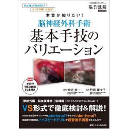
脳神経外科速報 2021年増刊号 脳神経外科手術 基本手技のバリエーション

出版社: メディカ出版
発行日: 2021-10-01
分野: 臨床医学:外科  >  雑誌
ISSN: 09171495
雑誌名:
特集: 脳神経外科手術 基本手技のバリエーション
電子書籍版: 2021-10-01 (電子書籍版)
書籍・雑誌
≪全国送料無料でお届け≫
取寄せ目安:8~14営業日

8,800 円(税込)

電子書籍
章別単位での購入はできません
ブラウザ、アプリ閲覧

8,800 円(税込)

目次

  • 特集 何が違って何が同じ? なぜその違いが出る? 本質が知りたい!
       脳神経外科手術 基本手技のバリエーション

    ◆1章 手術の基本コンセプトと技術
    ■A 手術教育
    〈総論1〉若手の教育(2 hands or 3 hands?)
    i.「2 hands派」の立場から
    ii.「3 hands派」の立場から─若手の顕微鏡下手術における指導的助手の役割

    ■B 開頭のバリエーション
    〈総論1〉前頭側頭開頭のバリエーション(適応,コンセプト,皮切デザイン,筋肉剥離)
    i. Pterional approachを企図した前頭側頭開頭
    ii.あらゆる病変に対応可能なアプローチとしての前頭側頭開頭法

    〈総論2〉後頭下開頭のバリエーション(適応,コンセプト,皮切デザイン,筋肉剥離)
    i. まず浅い術野の展開を
    ii.体位・頭部固定が最重要

    〈コラム1〉Orbitozygomatic(OZ)craniotomy
    〈コラム2〉2-layerのやり方:その工夫とメリット・デメリット
    i. Yasargil原法(1)
    ii.Yasargil原法2)

    ■C マイクロ操作
    〈総論1〉基本操作:シルビウス裂開放のバリエーション
    (シルビウス裂は広く開ける?必要最小限?/どこから入る?)
    i. 病変に応じたシルビウス裂開放
    ii.まずは十分なシルビウス裂開放から

    〈総論2〉器具の使い方のバリエーション
    (脳べらを積極的に使う?極力使わない?/吸引管,ハサミはどのように使い分ける?)
    i. 「脳べら積極派」の立場から
    ii.脳へらを極力使用しない手技

    〈総論3〉静脈温存のコンセプト
    (静脈はどこまで温存すべきか?/切断可能かどうかの判断法は?)
    i. 原則,すべての静脈を温存
    ii.術野展開の妨げとなる場合の対処法

    ■D アプローチ
    〈総論1〉前床突起切除法(intradural or extradural?/drill or CUSA?)
    i. 「Intraduralからの切除」の立場から
    ii.「Extradural派」の立場から

    〈総論2〉MCA, IC-PC動脈瘤のアプローチ
    (pterional or subfrontal?その適応とコンセプト/母血管の取り方)
    i. 「Pterional派」の立場から
    ii.「Subfrontal派」の立場から

    〈コラム1〉Dural ringの切開って?(適応,解剖,手技)─中枢側より剥離せよ


    ◆2章 頭部外傷の手術
    〈総論1〉頭部外傷手術のバリエーション
        (開頭術の適応あるいはタイミングは?/脳室ドレーンや硬膜下ドレーン挿入部から
         髄液漏れを防ぐための固定法のバリエーション/減圧開頭の範囲の決定法と
         術後CTでどう評価するか?/術中の止血困難な状況における対処法のバリエーション/
         HITTは行うべきか?/減圧開頭時の硬膜形成のバリエーション/
         ICPモニタリングの適応と留置部位に関する施設間のバリエーション)
    i. まず迅速な減圧.穿頭術も併用しつつ積極的な開頭術を実施
    ii.ICP亢進や意識レベル低下などの生理学徴候を優先して減圧開頭術を判断

    〈コラム1〉脳圧亢進に対する対処法─Multimodalなmonitoringを駆使せよ
    〈コラム2〉後頭下減圧開頭を行うための皮膚切開のバリエーション
    〈コラム3〉慢性硬膜下血腫に対する穿頭術で,術中にできる再発率低下のための工夫は?


    ◆3章 脳血管障害の手術
    ■A 頚動脈内膜剥離術(CEA)
    〈総論1〉内頚動脈側のプラーク処理(引き抜く?絶対に引き抜かない?)
    血管縫合時の運針作法(ロックをかける?かけない?/靴紐縫合する?しない?/
    intactな部分から縫う?縫わない?)
    i. 「引き抜かない派」のプラーク剥離・露出と血管縫合時の運針作法
    ii.CEAにおけるプラーク剥離と血管縫合

    〈コラム1〉CEAにおける血管遮断の工夫,遮断・遮断解除の順序,その本質について

    ■B 脳動脈瘤手
    〈総論1〉クリッピング
    (脳内に埋没した脳動脈瘤,完全に掘り起こす?十分なneckが出たらかける?)
    i. 「完全掘り起こし派」の立場から
    ii.「Neck派」の立場から

    〈総論2〉Acom動脈瘤へのアプローチのバリエーション
    (Pterional or interhemispheric approach?/視神経上面はどこまで剥離する?/
    半球間裂はどこまで開ける?/Rectal gyrusは吸う?)
    i. Pterional approachが第一選択
    ii.Interhemispheric approachとpterional approachの使い分け

    〈総論3〉クリップ選択のバリエーション
    (Temporary clipを使う?極力使わない?/Permanent clipの選択)
    i. 「Temporary clipを使用する派」として
    ii.Temporary clipを極力使用しない立場から

    〈コラム1〉くも膜下出血術中の脳圧コントロール
    〈コラム2〉低位内頚動脈系動脈瘤の母血管確保(方法,タイミング,その考え方)
    i. 頚部頚動脈確保, 前床突起削除, anterior petroclinoid ligamentの切開
    ii.原則,頚部での血管確保を

    ■C バイパス術
    〈総論1〉STA-MCAバイパスのコンセプト
    (皮膚切開はどうする?/ドナー剥離は動脈の表剥き?裏剥き?/Single or double bypass?/
     レシピエントは前頭葉?側頭葉?/間接吻合は置く?置かない?)
    ii. 間接バイパスのdonorとなる血管,硬膜,側頭筋などの組織の温存
    ii.血液や過剰な髄液流入のない術野をつくる

    〈コラム1〉STA graftを安全にはやくきれいに採取するには?


    ◆4章 脳腫瘍の手術
    〈総論1〉良性腫瘍の内減圧の方法のバリエーション・どう使い分けるか/
         腫瘍摘出中の止血法のバリエーション・どう使い分けるか/
         術中に綿片や脳べらを積極的に利用するか,しないか.どう利用するか
    i. くも膜を温存した剥離を
    ii.器具のセッティングは使用法と同様に重要

    〈総論2〉頭蓋底骨削除の方法(ハイスピードドリルまたは超音波手術器)
    i. 「ハイスピードドリル派」の立場から
    ii.「超音波手術器派」の立場から

    〈総論3〉グリオーマ摘出における各摘出器具の利点と使い分け(バイポーラまたはCUSA)
    i. 「吸引管とバイポーラ派」の立場から
    ii.「CUSA派」の立場から

    〈コラム1〉顕微鏡か内視鏡か外視鏡か,それぞれの利点と欠点は?
    ─頭蓋底腫瘍手術を念頭に
    〈コラム2〉術前腫瘍塞栓術(PTE)の適応─脳血管内治療医と腫瘍摘出術者の協同
    〈コラム3〉脳腫瘍手術における再手術時の留意点は?

この書籍の参考文献

参考文献のリンクは、リンク先の都合等により正しく表示されない場合がありますので、あらかじめご了承下さい。

本参考文献は電子書籍掲載内容を元にしております。

1章 手術の基本コンセプトと技術

P.19 掲載の参考文献
1) 出井勝ほか : 脳動脈瘤クリッピング術の技量の継承. 脳卒中の外科 37 : 192-6, 2009
2) 堤一生 : 私の手術教育-初心者指導の経験から. 脳卒中の外科 35 : 361-3, 2007
3) 吉金努ほか : 若手脳神経外科医が脳血管外科手術を習得するための教育法. 脳卒中の外科 47 : 33-40, 2019
4) 師井淳太ほか : 脳動脈瘤クリッピングの術者教育-秋田脳研「脳動脈瘤の外科コース」の成果と指導医のリスク管理について. 脳卒中の外科 42 : 422-6, 2014
P.29 掲載の参考文献
1) 出雲剛ほか : コロラドニードル, p.77 (飯原弘二編 : 脳神経外科診療プラクティス 2 脳神経外科の基本手技. 文光堂, 2014)
2) Poblete T, et al : Preservation of the nerves to the frontalis muscle during pterional craniotomy. J Neurosurg 122 : 1274-82, 2015
3) Yoshioka N, et al : Titanium mesh implant exposure due to pressure gradient fluctuation. World Neurosurg 119 : e734-9, 2018
4) Yasuhara T, et al : Japanese national questionnaire survey in 2018 on complications related to cranial implants in neurosurgery. Neurol Med Chir (Tokyo) 60 : 337-50, 2020
P.35 掲載の参考文献
1) Heros RC : Lateral suboccipital approach for vertebral and vertebrobasilar artery lesions. J Neurosurg 64 : 559-62, 1986
2) Bertalanffy H, et al : The dorsolateral, suboccipital, transcondylar approach to the lower clivus and anterior portion of the craniocervical junction. Neurosurgery 29 : 815-21, 1991
3) Bulsara KR, et al : Microsurgical management of 53 jugular foramen schwannomas : lessons learned incorporated into a modified grading system. J Neurosurg 109 : 794-803, 2008
4) Matsushima T, et al : Transcerebellomedullary fissure approach with special reference to methods of dissecting the fissure. J Neurosurg 94 : 257-64, 2001
5) Mussi AC, et al : Telovelar approach to the fourth ventricle : microsurgical anatomy. J Neurosurg 2 : 812-23, 2000
6) Matsushima T, et al : Microsurgical anatomy for lateral approaches to the foramen magnum with special reference to transcondylar fossa (supracondylar transjugular tubercle) approach. Skull Base Surgery 8 : 119-25, 1998
7) Matsushima K, et al : Partial transcondylar approach for ventral foramen magnum neurenteric cyst : 2-dimensional operative video. Oper Neurosurg (Hagerstown) 16 : E81, 2019
8) 河野道宏 : Transcondylar approach, 148-56 (佐伯直勝編 : 脳神経外科エキスパート 頭蓋底. 中外医学社, 東京, 2009)
9) 松島健ほか : 頚静脈孔腫瘍に対するアプローチ-特にS状静脈洞を温存する術式. 脳外速報 26 : 1041-7, 2016
10) Matsushima K, et al : Microsurgical and endoscopic anatomy for intradural temporal bone drilling and applications of the electromagnetic navigation system : various extensions of the retrosigmoid approach. World Neurosurg 103 : 620-30, 2017
11) 河野道宏 : Retrosigmoid approach, 110-21 (伊達勲編 : 新NS NOW 2 若手脳神経外科医が経験したい手術アプローチ. メジカルビュー社, 東京, 2015)
12) 河野道宏 : 聴神経腫瘍手術におけるトラブルへの対応. 脳外速報 24 : 24-32, 2014
13) Matsushima K, et al : Management of sigmoid sinus injury : retrospective study of 450 consecutive surgeries in the cerebellopontine angle and intrapetrous region. Oper Neurosurg (Hagerstown) 19 : 721-9. 2020
P.40 掲載の参考文献
1) 松島俊夫 : 後頭蓋窩3のルール, 1-6 (後頭蓋窩の微小解剖と手術. サイメッド・パブリケーションズ, 東京, 2006)
2) 小林広昌 : Q1-3, 16-21 (吉村紳一監修, 吉村幸司編集 : 専門医なら知っておきたい疾患・術式別 脳神経外科手術合併症の回避・対処法 Q&A 156. 脳外速報 2019 増刊 メディカ出版, 大阪, 2019)
3) 安部洋 : OA-PICA bypassを用いたVA・PICA動脈瘤手術. 脳外速報 29 : 1256-65, 2019
P.45 掲載の参考文献
1) Pellerin P, et al : Usefulness of the orbitofrontomalar approach associated with bone reconstruction for frontotemporosphenoid meningiomas. Neurosurgery 15 : 715-8, 1984
2) Hakuba A, et al : The orbitozygomatic infratemporal approach : a new surgical technique. Surg Neurol 26 : 271-6, 1986
3) Hakuba A, et al : A combined orbitozygomatic infratemporal epidural and subdural approach for lesions involving the entire cavernous sinus. J Neurosurg 71 : 699-704, 1989
4) McDermott MW, et al : Combined frontotemporal-orbitozygomatic approach for tumors of the sphenoid wing and orbit. Neurosurgery 26 : 107-16, 1990
5) Zabramski JM, et al : Orbitozygomatic craniotomy. Technical note. J Neurosurg 89 : 336-41, 1998
6) Alaywan M, Sindou M : Fronto-temporal approach with orbito-zygomatic removal. Surgical anatomy. Acta Neurochir (Wien) 104 : 79-83, 1990
7) 小寺俊昭ほか : Temporopolar approachによる脳底動脈分岐部動脈瘤クリッピング術. 脳外速報 21 : 846-5, 2011
8) Aziz KMA, et al : The one-piece orbitozygomatic approach : the MacCarty burr hole and the inferior orbital fissure as keys to technique and application. Acta Neurochir (Wien) 144 : 15-24, 2002
9) Tanriover N, et al : One-piece versus two-piece orbitozygomatic craniotomy : quantitative and qualitative considerations. Neurosurgery 58 (4 Suppl 2) : 229-37, 2006
10) Ohata K, et al : "Orbitozygomatic infratemporal approach", 1-35〔Hakuba A (ed) : Surgical anatomy of the skull base. Miwa shoten, Tokyo, 1996〕
11) Kodera T, et al : Orbitozygomatic craniotomy with modified Zabramski's technique : a technical note and anatomic and clinical findings. World Neurosurg 97 : 49-57, 2017
12) 小寺俊昭 : "Orbitozygomatic approachの開頭", 107-14 (菊田健一郎編 : 脳神経外科手術スキルアップガイド. 中外医学社, 東京, 2015)
13) Martins C, et al : Role of the zygomaticofacial foramen in the orbitozygomatic craniotomy : anatomic report. Neurosurgery 53 : 168-73, 2003
P.48 掲載の参考文献
1) Yasargil MG : Microneurosurgery Vol I, Georg Thieme Verlag, Stuttgart, New York, 1984, 215-25
2) 原貴行 : 頭皮の外科解剖と皮弁のデザイン, 54-60 (飯原弘二編 : 脳神経外科診療プラクティス 2 脳神経外科の基本手技, 文光堂, 東京, 2014)
P.54 掲載の参考文献
1) 瀧澤克己 : 前頭側頭開頭, 14-25 (寺本明担当編集 : 整容脳神経外科Update, NS NOW No. 13. メジカルビュー社, 東京, 2011)
P.64 掲載の参考文献
1) 田中雄一郎ほか : Pterional approach (蝶形骨縁到達法) : ピットフォール回避の知識. 脳外誌 19 : 727-32, 2010
2) Mori K, et al. Keyhole strategy aiming at minimizing hospital stay for surgical clipping of unruptured middle cerebral artery aneurysms. J Neurosurg 130 : 1359-66, 2019
3) 石黒太一ほか : 前方循環未破裂脳動脈瘤に対する低侵襲化直達手術. No Shinkei Geka 48 : 1005-12, 2020
4) 谷川緑野ほか : 内頚動脈瘤手術におけるdistal transsylvian approachのアドバンテージと技術的ポイント. 脳卒中の外 32 : 19-24, 2004
5) 竹田理々子ほか : 前交通動脈瘤に対するpterional approach. 脳外速報 22 : 744-54, 2012
6) Ture U, et al : Topographic anatomy of the insular region. No Shinkei Geka 90 : 720-33, 1999
7) Roblot P, et al : Skin landmarks to main cerebral structures : how to identify the main cerebral sulci? An anatomical study. Surg Radiol Anat 2021 May 1. [Online ahead of print.]
8) Ribas GC, et al : The anterior sylvian point and the suprasylvian operculum. Neurosurg Focus 18 : E2, 2005
9) Lee SH, et al : The normal angiographic sylvian point on the lateral cerebral angiogram. Neuroradiology 17 : 101-3, 1979
10) Kazumata K, et al : Operative anatomy and classification of the sylvian veins for the distal transsylvian approach. Neurol Med Chir (Tokyo) 43 : 427-33, 2003
11) 石川達哉 : 脳動脈瘤手術におけるアプローチとスペースの作り方 : 超初心者のためのシルビウス裂開放の考え方. 脳外速報 19 : 26-36, 2009
12) 杉山拓 : はじめてのシルビウス裂の開放. 脳外速報 21 : 1338-46, 2011
13) 大里俊明ほか : Pterional approachにおけるシルビウス裂開放のコツ. 脳卒中の外科 31 : 43-6, 2003
14) 波出石弘ほか : 脳動脈瘤手術の工夫. 脳卒中の外科 34 : 340-6, 2006
15) Matano F, et al : Olfactory preservation during anterior interhemispheric approach for anterior skull base lesions : technical note. Neurosurg Rev 39 : 63-9, 2016
P.68 掲載の参考文献
1) 上山博康ほか : 脳動脈瘤手術 基本技術とその応用. 南江堂, 東京, 354p, 2010.
P.74 掲載の参考文献
1) 横山和弘ほか : 脳ベラを用いない内頚動脈瘤の手術手技. 脳卒中の外科 31 : 279-84, 2003
2) 横山和弘ほか : 未破裂脳動脈瘤直達手術におけるpterional approachの無理のない術野拡大の工夫. 脳卒中の外科 39 : 84-8, 2011
3) 水谷徹 : 無欠の術野を目指す脳動脈瘤手術の基本-まずは手の安定と, ハサミ, 吸引管の基本的な使い方. 脳外速報 24 : 1064-72, 2014
4) 水谷徹ほか : 安全確実なclip操作を意識した未破裂脳動脈瘤手術-Blading techniqueのススメ. 脳外誌 24 : 822-32, 2015
5) 水谷徹 : 動脈瘤周囲の剥離と安全確実なクリップのかけ方は? No Shinkei Geka 49 : 52-71, 2021
6) 水谷徹 : 解離性脳動脈瘤への探求心 脳動脈瘤手術の極意, 工夫. 脳外速報 29 : 348-58, 2019
P.86 掲載の参考文献
1) Kazumata K, et al : Operative anatomy and classification of the sylvian veins for the distal transsylvian approach. Neurol Med Chir (Tokyo) 43 : 427-33, 2003
2) 赤須功ほか : シルビウス裂深部の正常異形静脈 (common vertical trunk) の存在はshort M1 中大脳動脈clipping術に伴う虚血性合併症率を高める. 脳卒中の外科 49 : 174-9, 2021
3) Maekawa H, et al : Venous-preserving sylvian dissection. World Neurosurgery 84 : 2043-52, 2015
4) 斉藤文男ほか : Pterional approachにおける静脈処理の問題点-Fronto-temporal bridging veinの考察. 脳卒中の外科 26 : 237-41, 1998
5) Ferroli P, et al : Venous sacrifice in neurosurgery : new insights from venous indocyanine green videoangiography. J Neurosurg 115 : 18-23, 2011.
6) Komiyama M : Functional venous anatomy of the brain for neurosurgeons. 脳外誌 26 : 488-95, 2017
7) 小宮山雅樹 : 脳静脈の機能解剖. 脳外誌 18 : 821-9, 2009
P.92 掲載の参考文献
1) 久須美真理ほか : IC-PC大型動脈瘤に対する前床突起切除-適応, 方法, 注意点-. 脳卒中の外科 42 : 288-93, 2014
P.97 掲載の参考文献
1) Noguchi A, et al : Extradural anterior clinoidectomy. Technical note. J Neurosurg 102 : 945-50, 2005
2) Barnett SL, et al. : Intradural clinoidectomy and postoperative headache in patients undergoing aneurysm surgery. Neurosurgery 67 : 906-10, 2010
P.102 掲載の参考文献
1) Yasargil MG, et al : The microsurgical approach to intracranial aneurysms. Surg Neurol 3 : 7-14, 1975
2) 上山博康ほか : 脳動脈瘤手術-基本技術とその応用. 南江堂, 東京, 354p, 2010
P.107 掲載の参考文献
1) Lehecka M, et al. Helsinki microneurosurgery basics and tricks. Druckerei Hohl GmbH et Co. KG, Balgheim, 2011
2) Yamahata H, et al : Efficacy and safety of the pterional keyhole approach for the treatment of anterior circulation aneurysms. Neurosurg Rev 37 : 629-36, 2014
P.113 掲載の参考文献
1) 坪井俊之ほか : Paraclinoid aneurysmに対するDolenc's approach-より安全に前床突起削除を行うために. 脳外速報 25 : 692-703, 2015
2) 京島和彦ほか : 内頚動脈硬膜輪近傍の外科解剖. 130-6 (顕微鏡下手術のための脳神経外科解剖 V, 第6回微小脳神経外科解剖セミナー講演集, 1992)
3) Rhoton AL, et al : Chapter 2 Neural structures. 35 (The orbit and sellar region : microsurgical anatomy and operative approaches. George Thieme Verlag, Stuttgart, 1996)

2章 頭部外傷の手術

P.120 掲載の参考文献
1) 頭部外傷治療・管理のガイドライン作成委員会編 : 頭部外傷治療・管理のガイドライン 第4版. 医学書院, 東京, 2019
2) Suehiro E, et al : Predictors of deterioration indicating a requirement for surgery in mild to moderate traumatic brain injury. Clin Neurol Neurosurg 127 : 97-100, 2014
3) Kawamata T, et al : Tissue hyperosmolality and brain edema in cerebral contusion. Neurosurg Focus 22 : E5, 2007
4) Matsushima K, et al : Emergent operation for isolated severe traumatic brain injury : Does time matter? J Trauma Acute Care Surg 79 : 838-42, 2015
5) Mendelow AD, et al : Early surgery versus initial conservative treatment in patients with traumatic intracerebral hemorrhage (STITCH [Trauma]) : the first randomized trial. J Neurotrauma 32 : 1312-23, 2015
6) Jiang JY, et al : Efficacy of standard trauma craniectomy for refractory intracranial hypertension with severe traumatic brain injury : a multicenter, prospective, randomized controlled study. J Neurotrauma 22 : 623-8, 2005
7) Kiyohira M, et al : Combined strategy of burr hole surgery and elective craniotomy under intracranial pressure monitoring for severe acute subdural hematoma. Neurol Med Chir (Tokyo) 61 : 253-9, 2021
8) Hawryluk GWJ, et al : A management algorithm for patients with intracranial pressure monitoring : the Seattle International Severe Traumatic Brain Injury Consensus Conference (SIBICC). Intensive Care Med 45 : 1783-94, 2019
P.127 掲載の参考文献
1) 萩原靖 : ICPモニタリングによる脳神経救急管理の実際. 脳外速報 29 : 939-47, 2019
2) 頭部外傷治療・管理のガイドライン作成委員会編 : 頭部外傷治療・管理のガイドライン 第4版. 医学書院, 東京, 2019
3) Miabi Z, et al : Delineation of lateral tentorial sinus with contrast-enhanced MR imaging and its surgical implications. AJNR 25 : 1181-8, 2004
4) 吉岡伸高ほか : ゴアテックス人工硬膜感染症の治療方針. 脳外誌 28 : 19-25, 2019
5) 萩原靖 : トップナイフの閃き-頭部を切る・開ける : 重症頭部外傷における緊急開頭の決断. 救急医学 44 : 1866-75, 2020
P.131 掲載の参考文献
1) Andrew IR et al : Current recommendations for neurotrauma. Curr Opin Crit Care 6 : 281-92, 2000
2) Carney N, et al : Guideleines for the management of severe traumatic brain injury, fourth edition. Neurosurgery 80 : 6-15, 2017
3) 頭部外傷治療・管理のガイドライン作成委員会 : 頭部外傷治療・管理のガイドライン, 第4版. 医学書院, 東京, 2019
4) Kiyohira M, et al. Combined strategy of burr hole surgery and elective craniotomy under intracranial pressure monitoring for severe acute subdural hematoma. Neurol Med Chir (Tokyo) 61 : 253-9, 2021
5) Okonkwo D, et al : Brain tissue oxygen monitoring and management in severe traumatic brain injury (BOOST-II) : a phase II randomized trial. Crit Care Med 45 : 1907-14, 2017
6) 横堀將司ほか : 頭部外傷の病態と治療. 日医大医会誌 15 : 71-9, 2019
P.136 掲載の参考文献
1) 太田富雄 : 10章 頭部外傷 7. 外傷性頭蓋内血腫, 1116-9 (松谷雅生ほか編 : 脳神経外科学 改訂第13版, 金芳堂, 京都, 2021)
2) Zuccarello M, et al : Epidural hematomas of the posterior cranial fossa. Neurosurg 8 : 434-7, 1981
3) Roth C, et al : Does prone positioning increase intracranial pressure? A retrospective analysis of patients with acute brain injury and acute respiratory failure. Neurocrit Care 21 : 186-91, 2014
4) Teranishi Y, et al : Determination of the keyhole position in a lateral suboccipital retrosigmoid approach. Neurol Med Chir (Tokyo) 54 : 261-6, 2014
P.140 掲載の参考文献
1) Hirai S, et al : Postoperative recurrence of chronic subdural hematoma is more frequent in patients with blood type A. J Neurosurg 2021 Jan 1〔Online ahead of print〕
2) Mori K, et al : Surgical treatment of chronic subdural hematoma in 500 consecutive cases : clinical characteristics, surgical outcome, complications, and recurrence rate. Neuro Med Chir (Tokyo) 41 : 371-81, 2001
3) 高山東春ほか : 慢性硬膜下血腫の再発因子に関する後ろ向き統計学的分析 : 単変量, 多変量解析による検討. No Shinkei Geka 40 : 871-6, 2012
4) Edlmann E, et al : Pathophysiology of chronic subdural haematoma : inflammation, angiogenesis and implications for pharmacotherapy. J Neuroinflammation 14 : 108, 2017
5) Toi H, et al : Determining if cerebrospinal fluid prevents recurrence of chronic subdural hematoma : a multi-center prospective randomized clinical Trial. J Neurotrauma 36 : 559-64, 2019

3章 脳血管障害の手術

P.146 掲載の参考文献
1) 一ノ瀬努ほか : 高度石灰化病変に対する確実な顕微鏡下頚動脈内膜剥離術-内膜内剥離による段階的切除と病理学的考察-. 脳卒中の外科 46 : 429-34, 2018
2) Ichinose T, et al : Microsurgical interintimal dissection in carotid endarterectomy. World Neurosurg 82 (1-2) : e225-8, 2013
P.150 掲載の参考文献
1) 宇野昌明 : 頚動脈内膜剥離術. 脳外誌 24 : 840-5, 2015
2) 吉野正紀ほか : 安全にCEAを施行するために-手術手技, 周術期管理, 術者養成の検討-. 脳卒中の外科 39 : 257-61, 2011
3) 一ノ瀬努ほか : 高度石灰化病変に対する確実な顕微鏡下頚動脈内膜剥離術-内膜内剥離による段階的切除と病理学的考察-. 脳卒中の外科 46 : 429-34, 2018
4) 宇野昌明 : 頚動脈内膜剥離術. 脳外誌 21 : 943-8, 2012
P.154 掲載の参考文献
1) Verhoeven BA, et al : Carotid atherosclerotic plaque characteristics are associated with microembolization during carotid endarterectomy and procedural outcome. Stroke 36 : 1735-40, 2005
2) Izumo T, et al : The orthodox techniques of carotid endarterectomy. Neurosurgical Emergency 22 : 93-96, 2017
3) Jansen C, et al : Impact of microembolism and hemodynamic changes in the brain during carotid endarterectomy. Stroke 25 : 992-7, 1994
P.158 掲載の参考文献
石川達哉 : 逆境を乗り越えるためのリクツとワザ, 190-6 (石川達哉編 : 新版クリッピング・バイパス・CEA のリクツとワザ-脳血管外科の学び方・教え方. メディカ出版, 大阪, 2019)
坪井俊之ほか : 合併症ZERO を目指した動脈瘤クリッピング術-理想的なclosure lineを意識した穿通枝の剥離温存の工夫. 脳外速報 29 : 1266-75, 2019)
P.162 掲載の参考文献
1) Kimura T, et al. Preoperative evaluation of unruptured cerebral aneurysms by fast imaging employing steady-state acquisition image. Neurosurgery 69 : 412-9, 2011
2) Sakuma J, et al. Monitoring and preventing blood flow insufficiency due to clip rotation after the treatment of internal carotid artery aneurysms : technical note. J Neurosurg 100 : 960-2, 2004
P.167 掲載の参考文献
1) Fujiwara H, Yasui N, Nathal-Vera E, et al. Anosmia after anterior communicating artery aneurysm surgery : comparison between the anterior interhemispheric and basal interhemispheric approaches. Neurosurgery 38 : 325-8, 1996
P.181 掲載の参考文献
1) Dujovny M, et al : Minimum vascular occlusive force. J Neurosurg 51 : 662-8, 1979
2) 石川達哉 : 追い込むtemporary clip, 追い込まれたtemporary clip-脳動脈瘤におけるtemporary clipの使い方 : 後編. 脳外速報 22 : 1004-10, 2012
3) Samson D, et al : A clinical study of the parameters and effects of temporary arterial occlusion in the management of intracranial aneurysms. Neurosurgery 34 : 22-9, 1994
4) Kobayashi S, et al : Quantitative cerebral perfusion assessment using microscope-integrated analysis of intraoperative indocyanine green fluorescence angiography versus positron emission tomography in superficial temporal artery to middle cerebral artery anastomosis. Surg Neurol Int 5 (Supplement) : 135, 2014
5) Ishikawa T, et al : Concept of ideal closure line for clipping of middle cerebral artery aneurysms : technical note. Neurol Med Chir (Tokyo) 49 : 273-7, 2009
P.186 掲載の参考文献
1) 水谷徹ほか : 大型/巨大中大脳動脈瘤の直達クリッピング. 脳外速報 28 : 560-72, 2018
2) 石川達哉 : 追い込むtemporary clip, 追い込まれたtemporary clip-脳動脈瘤手術のおけるtemporary clipの使い方 : 前編-. 脳外速報 22 : 878-85, 2012
3) 石川達哉 : 追い込むtemporary clip, 追い込まれたtemporary clip-脳動脈瘤手術のおけるtemporary clipの使い方 : 後編-. 脳外速報 22 : 1004-9, 2012
4) 永田和哉 : ネックの形状を意識したクリッピング術-クリップ選択の基本的な考え方-. 脳外速報 15 : 328-34, 2005
5) 澤田元史ほか : 未破裂脳動脈瘤手術のピットホール-動脈硬化合併例での問題点と手術対策-. 脳卒中の外科 38 : 348-52, 2010
6) 水谷徹 : 動脈瘤周囲の剥離と安全確実なクリップのかけ方は? No Shinkei Geka 49 : 52-71, 2021
7) 水谷徹ほか : 安全確実なclip操作を意識した未破裂脳動脈瘤手術-Blading techniqueのススメ-. 脳外誌 24 : 822-32, 2015
8) 水谷徹 : 無血の術野を目指す脳動脈瘤手術の基本-両手の同時操作で最後までclip先端を見届ける : Bladingテクニックのススメ-. 脳外速報 25 : 128-38, 2015
P.190 掲載の参考文献
1) Paine JT, et al : Intraoperative ventricular puncture. Neurosurgery 22 : 1107-9, 1988
2) Park J, et al : Revision of Paine's technique for intraoperative ventricular puncture. Surg Neurol 70 : 503-8, 2008
3) Hyun SJ, et al : Novel entry point for intraoperative ventricular puncture during the transsylvian approach. Acta Neurochir (Wien) 149 : 1049-51, 2007
4) Matsuzaki H, et al : Use of Paine's technique : projecting puncture point to the skull and skin. World Neurosurgery 104 : 45-7, 2017
5) Moon HH, et al : Brain injuries during intraoperative ventriculostomy in the aneurysmal subarachnoid hemorrhage patients. J Korean Neurosurg Soc 46 : 215-20, 2009
P.193 掲載の参考文献
1) Niibo T, et al : Prediction of the difficulty of proximal vascular control using 3D-CTA for the surgical clipping of internal carotid artery-posterior communicating artery aneurysms. J Neurosurg 2020 Apr 10 : 1-8. [Online ahead of print.]
2) Kim JH, et al : Simple anterior petroclinoid fold resection in the treatment of low-lying internal carotid-posterior communicating artery aneurysms. Surg Neurol 72 : 142-5, 2009
3) Tanaka Y, et al : Protective dural flap for bone drilling at the paraclinoid region and porus acusticus : technical note. Neurol Med Chir (Tokyo) 43 : 416-8, 2003
P.197 掲載の参考文献
1) Ota N, et al : Predictor of visual impairment following paraclinoid aneurysm surgery : Special consideration of surgical microanatomy related to paraclinoid structures. Oper Neurosurg (Hagerstown) 20 : 45-54, 2020
2) Takeuchi S, et al : Retrograde suction decompression for clip occlusion of internal carotid artery communicating segment aneurysms. World Neurosurg 89 : 19-25, 2016
P.203 掲載の参考文献
1) Saito N, et al : Insights on the revascularization mechanism for treatment of moyamoya disease based on the histopathologic concept of angiogenesis and arteriogenesis. World Neurosurg 75 : 204-5, 2011
2) Heil M, et al : Arteriogenesis versus angiogenesis : similarities and differences. J Cellilar and Molecular Medicine 10 : 45-55, 2006
P.208 掲載の参考文献
2) Funaki T, et al : High rebleeding risk associated with choroidal collateral vessels in hemorrhagic moyamoya disease : analysis of a nonsurgical cohort in the Japan Adult Moyamoya Trial. J Neurosurg 130 : 337-673, 2019
3) Funaki T, et al : The targeted bypass strategy for preventing hemorrhage in moyamoya disease : technical note. Neurol Med Chir (Tokyo) 59 : 517-22, 2019
P.212 掲載の参考文献
1) Powers WJ, et al : Extracranial-intracranial bypass surgery for stroke prevention in hemodynamic cerebral ischemia : the Carotid Occlusion Surgery Study randomized trial. JAMA 306 : 1983-92. 2011
2) 村井保夫ほか : 血管吻合の基本手技 (森田明夫担当編集 : バイパス術のすべて 新NS Now 1 メジカルビュー社, 東京, 2015)
3) Kamiyama H, et al : Visualization of the ostium of an arteriotomy in bypass surgery. Neurosurgery 33 : 1109-10, 1993
4) Murai Y, et al : The feasibility of detecting cerebral blood flow direction using the indocyanine green video angiography. Neurosurg Rev 39 : 685-90, 2016

4章 脳腫瘍の手術

P.219 掲載の参考文献
1) Nagahama Y, et al : Localized controlledfibrin glue application with gelatin sponge forhemostasis and dural defect repair : Technical note. Interdisciplinary Neurosurgery 18 : 100476, 2019
P.226 掲載の参考文献
1) Fukushima T, et al [eds] : Fukushima Manual of Skull Base Dissection Third edition. AF Neurovideo, Raleigh, NC, 2010
2) 森田明夫 : 良性腫瘍の手術適応と取り方, 94-110 (木内博之ほか監修, 河野道宏編集 : プライム脳神経外科 5 頭蓋底腫瘍. 三輪書店, 東京, 2020)
3) Zomorodi A, et al : Two surgeons four-hand microneurosurgery with universal holder system : technical note. Neurosurg Rev 40 : 523-6, 2017
4) 河野道宏 : 聴神経腫瘍手術-手術適応と機能温存-. 脳外誌 23 : 20-8, 2014
5) Nonaka Y, et al : Micropatties are indispensable instrument for successful microneurosurgery ; Technical note. World Neurosurg 133 : 60-5, 2019
P.232 掲載の参考文献
1) Kondo S, et al : Thermological study of drilling bone tissue with a high-speed drill. Neurosurgery 46 : 1162-8, 2000
3) Siljander BR, et al : Cool Mist Irrigation Improves Heat Dissipation during Surgical Bone Drilling. J Neurol Surg B Skull Base 75 : 243-6, 2014
4) Wanibuchi M, et al : Hearing preservation surgery for vestibular schwannomas via the retrosigmoid transmeatal approach : surgical tips. Neurosurg Rev 37 : 431-44 ; discussion 444, 2014
5) Wanibuchi M, et al : Training for Skull Base Surgery with a Colored Temporal Bone Model Created by Three-Dimensional Printing Technology. World Neurosurg 91 : 66-72, 2016
6) Wanibuchi M, et al : Skull base training and education using an artificial skull model created by selective laser sintering. Acta Neurochir (Wien) 152 : 1055-9 ; discussion 1059-60, 2010
P.236 掲載の参考文献
1) Hadeishi H, et al : Anterior clinoidectomy and opening of the internal auditory canal using an ultrasonic bone curette. Neurosurgery 52 : 867-71, 2003
P.240 掲載の参考文献
1) 齋藤太一ほか : グリオーマの再手術, 68-79 (森田明夫ほか編 : 新NS NOW 16 Neurosurgical Re-Operations脳神経外科における再手術・再治療. メジカルビュー社, 東京, 2018)
2) 村垣善浩ほか : 膠芽腫, 118-23 (河本圭司ほか編 : イラストレイテッド 脳腫瘍外科学. 医学書院, 東京, 2011)
3) 齋藤太一ほか : Insular gliomaの手術, 146-56 (杉山一彦ほか編 : 悪性脳腫瘍のすべて-Neuro-Oncologyの教科書-遺伝子診断時代の臨床リアルワールド. メディカ出版, 大阪, 2020)
P.250 掲載の参考文献
1) Rosler J et al : Clinical implementation of a 3D4K-exoscope (Orbeye) in microneurosurgery. Neurosurg Rev : 2021 Jun 18 [Epub ahead of print]
2) Hong CS et al : Comparison of endoscope versus microscope-assisted resection of deep-seated intracranial lesions using a minimally invasive port retractor system. J Neurosurg 124 : 799-810, 2016
3) 阿久津博義 : 神経内視鏡における4 hand surgeryのセッティングとコツ, 24-33 (伊達勲編 : 新NS NOW 19 Advanced 神経内視鏡手術 神経内視鏡の可能性をさらに拡げる. メジカルビュー社, 東京, 2019)
P.255 掲載の参考文献
1) Przybylowski CJ, et al : Preoperative embolization versus no embolization for WHO grade I intracranial meningioma : a retrospective matched cohort study. J Neurosurg 27 : 1-8, 2020
2) Wanibuchi M, et al : Quantitative assessment of flow reduction after feeder embolization in meningioma by using pseudocontinuous arterial spin labeling. World Neurosurg 93 : 237-45, 2016
3) Asai K, et al : Efficacy of endovascular intratumoral embolization for meningioma : assessment using dynamic susceptibility contrast-enhanced perfusion-weighted imaging. J Neurointerv Surg Mar 15, 2021 [Online ahead of print]
4) Mayercik V, et al : Arterial spin-labeling MRI identifies hypervascular meningiomas. AJR Am J Roentgenol 213 : 1124-8, 2019
5) 藤本康倫, 他 : 髄膜腫に対する術前塞栓術-塞栓効果評価と病理診断-. Neuro-Oncologyの進歩 26 : 1-11, 2019
6) Bendszus M, et al : Is there a benefit of preoperative meningioma embolization? Neurosurgery 47 : 1306-11, 2000
7) Brandel MG, et al : Impact of preoperative endovascular embolization on immediate meningioma resection outcomes. Neurosurg Focus 44 : E6, 2018
8) Ampie L, et al : Safety and outcomes of preoperative embolization of intracranial hemangioblastomas : A systematic review. Clin Neurol Neurosurg 150 : 143-51, 2016
9) Imai T, et al : Successful preoperative endovascular embolization of an extreme hypervascular glioblastoma mimicking an arteriovenous malformation. World Neurosurg 86 : 512. e1-4, 2016
10) 高橋和孝, 他 : 血管内治療と二期的摘出術が有用であった大型・hypervascular聴神経腫瘍の1手術例. 脳外誌 24 : 544-50, 2015
P.261 掲載の参考文献
1) Nanda A, et al : Relevance of Simpson grading system and recurrence-free survival after surgery for World Health Organization Grade I meningioma. J Neurosurg 126 : 201-11, 2017
2) Winther TL, et al : Significance of the extent of resection in modern neurosurgical practice of World Health Organization grade I meningiomas. World Neurosurg 99 : 104-10, 2017
3) Magill ST, et al : Surgical outcomes after reoperation for recurrent skull base meningiomas. J Neurosurg 130 : 876-83, 2019
4) Behari S, et al : Surgical strategies for giant medial sphenoid wing meningiomas : a new scoring system for predicting extent of resection. Acta Neurochir (Wien) 150 : 865-77, 2008
5) Bir SC, et al : Comparison of the surgical outcome of pterional and frontotemporal-orbitozygomatic approaches and determination of predictors of recurrence for sphenoid wing. World Neurosurg 99 : 308-19, 2017
6) Pirotte BJM, et al : Lateral and Middle Sphenoid Wing Meningiomas, 371-8 (Lee JH (ed) : Meningiomas. Springer, London, 2009)
7) Mansouri A, et al : Surgically resected skull base meningiomas demonstrate a divergent postoperative recurrence pattern compared with non-skull base meningiomas. J Neurosurg 125 : 431-40, 2016
8) Ouyang T, et al : Sphenoid wing meningiomas : Surgical strategies and evaluation of prognostic factors influencing clinical outcomes. Clin Neurol Neurosurg 134 : 85-90, 2015

最近チェックした商品履歴

Loading...