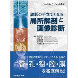
読影の手立てとなる局所解剖と画像診断
商品紹介
実質臓器に比べてこれまであまり注目されてこなかったCana(l 管)・Foramen(孔)・Fissure(裂)・Space(腔)およびMembrane(膜)などの解剖上の構造物に焦点をあて,構造物がどこに位置するのか、どのような意味を持つ構造物なのか、その画像診断上の意義を徹底解説。各領域のエキスパートが構造物を押さえてどう読影しているのかよくわかるよう,充実したシェーマと画像により細かいポイントまで記載。読影と診断の技能をより高めるために必携の一冊。
目次
- 第1章 頭頸部 Head & Neck
Overview—頭蓋底の解剖 Anatomy of the skull base
Foramen cecum 盲孔
Foramen of cribriform plate 篩板孔
Optic canal 視神経管
Superior orbital fissure 上眼窩裂
Inferior orbital fissure 下眼窩裂
Pterygopalatine fossa 翼口蓋窩
Foramen rotundum 正円孔
Foramen ovale 卵円孔
Carotid canal 頸動脈管
Foramen lacerum 破裂孔
Facial canal 顔面神経管
Internal auditory canal 内耳道
Inferior tympanic canaliculus 下鼓室小管
Foramen magnum 大後頭孔
Jugular foramen 頸静脈孔
Hypoglossal canal 舌下神経管
Pterygoid canal 翼突管
Foramen spinosum 棘孔
Foramen Vesalius Vesalius孔
Condylar canal 顆管
Overview—頭頸部の解剖 Anatomy of the fascia & space
Pharyngobasilar fascia 咽頭頭底筋膜
Parapharyngeal space 傍咽頭間隙
Tensor-vascular-styloid fascia(TVSF)
Parotid space 耳下腺間隙
Infratemporal fossa 側頭下窩
Masticator space 咀嚼筋間隙
Buccal space 頬間隙
Sublingual space 舌下間隙
Submandibular space 顎下間隙
Carotid sheath 頸動脈間隙
Retropharyngeal space 咽頭後間隙
Danger space 危険間隙
Perivertebral space 椎周囲間隙
Pharyngeal mucosal space 咽頭粘膜間隙
Visceral space 臓器間隙
Peritonsillar space 扁桃周囲間隙
Posterior cervical space 後頸間隙
Preepiglottic space,paraglottic space 前喉頭蓋間隙,傍声帯間隙
第2章 胸部 Chest & Heart, Great Vessel
Overview—胸部の解剖 Anatomy of the chest
Anterior junction line 前接合線
Posterior junction line 後接合線
Azygoesophageal recess 奇静脈食道陥凹
Left border of descending thoracic aorta 胸部下行大動脈線(左縁)
Left paraspinal line 左傍脊椎線
Right paratracheal stripe 右傍気管線
Preaortic recess 前大動脈陥凹
Aortopulmonary window 大動脈肺動脈窓
Retrosternal space 胸骨後腔
Retrocardiac space 心臓後腔
Kerley's lines カーリー線
Major fissure 大葉間裂(superomedial major fiisure,
superolateral major fissure, vertical fissure line)
Minor fissure 小葉間裂
Accessory fissure 副葉間裂:上副葉間裂,下副葉間裂,左小葉間裂,奇静脈裂
Pulmonary ligament 肺靭帯(肺間膜)
Overview—心膜の解剖 Anatomy of the pericardium
Transverse pericardial recess 心膜横洞
Oblique pericardial recess 心膜斜洞
Superior pericardial recess・left lateral pulmonic recess 上心膜腔・左外側陥凹
第3章 腹部・骨盤部 Abdominal and Pelvic region
Overview―腹膜腔の解剖 Anatomy of the intra-peritoneal space
Subphrenic space 横隔膜下腔
Right subhepatic space 右肝下腔
Paracolic gutter 傍結腸溝
Lesser sac 網嚢
Round ligament 肝円索
Hepatoduodenal ligament 肝十二指腸間膜
Gastrohepatic ligament 肝胃間膜
Gastrocolic ligament 胃結腸間膜
Gastrosplenic ligament 胃脾間膜
Ligament of Treitz Treitz靭帯
Transverse mesocolon 横行結腸間膜
Sigmoid mesocolon S状結腸間膜
Small bowel mesentery 小腸間膜
Overview―後腹膜腔の解剖 Anatomy of the retroperitoneal space
Anterior pararenal space 前腎傍腔
Perirenal space 腎周囲腔
Posterior pararenal space 後腎傍腔
Overview―骨盤部腹膜外腔の解剖 Anatomy of the intra-and extra-pelvic spaces
Prevesical space(retropubic space, Retzius space) 膀胱前腔
Perivesical space 膀胱周囲腔
Perirectal space 直腸周囲腔
Presacral space 仙骨前腔
Inguinal canal 鼡径管
Femoral canal 大腿管
Obturator foramen 閉鎖孔
Greater sciatic foramen 大坐骨孔
第4章 骨・関節・軟部 Bones, joints & soft tissue
Overview―肩の解剖 Anatomy of the shoulder joint
Acromio-humeral interval 肩峰骨頭間距離
Rotator interval 腱板疎部
Quadrilateral space 四辺形間隙,外側四角腔
Suprascapular notch, spinoglenoid notch 肩甲上切痕,棘下切痕
Glenohumeral ligament (関節上腕靱帯 上・中・下を含む)肩関節腔
Overview―肘の解剖 Anatomy of the elbow joint
Cubital tunnel 肘部管
Radial tunnel 橈骨神経管
Capitellar fat pad,trochlear fat pad 上腕骨小頭脂肪体,上腕骨滑車部脂肪体
Synovial fold(elbow) 滑膜ヒダ(肘)
Overview―手関節の解剖 Anatomy of the wrist joint
Carpal tunnel 手根管
Guyon canal Guyon管
Extensor compartments of the wrist 指伸筋のコンパートメント
Overview―股関節の解剖 Anatomy of the hip joint
Lessor sciatic notch 小坐骨切痕
Obturator sulcus 閉鎖溝
Piriform fossa 梨状窩
Ischiofemoral interval 坐骨大腿間距離
Overview―膝の解剖 Anatomy of the knee
Hoffa fat pad Hoffa脂肪体,膝蓋下脂肪体
Synovial plica(knee) 滑膜ヒダ(膝)
Compartment of medial collateral ligament 内側側副靭帯で分けられるコンパートメント
Meniscofemoral ligament,meniscotibial ligament 半月大腿靱帯,半月脛骨靱帯
Pes anserinus 鵞足
Iliotibial band 腸脛靱帯
Overview―足関節の解剖 Anatomy of the ankle joint
Tarsal tunnel 足根管
Anterior tarsal sinus 前足根管
Sinus tarsi 足根洞
Kager fat pad Kager脂肪体
この書籍の参考文献
参考文献のリンクは、リンク先の都合等により正しく表示されない場合がありますので、あらかじめご了承下さい。
本参考文献は電子書籍掲載内容を元にしております。
第1章 頭頸部 Head & Neck
P.16 掲載の参考文献
P.18 掲載の参考文献
1) Lewinska-Smiaek B, et al:Anatomy of the adult foramen caecum. Eur J Anat, 17:142-145, 2013.
2) Boyd GI:The emissary foramina of the cranium in man and the athropoids. J Anat, 65:108-121, 1929.
P.21 掲載の参考文献
P.23 掲載の参考文献
1) 久徳茂雄:顔面外傷のblow out fractureと視神経管骨折の治療. 痛みと臨床, 5:130-136. 2005.
2) Kornreich L, et al:Optic pathway glioma:correlation of imaging findings with the presence of neurofibromatosis. AJNR Am J Neuroradiol, 22:1963-1969, 2001.
P.24 掲載の参考文献
P.26 掲載の参考文献
P.29 掲載の参考文献
1) Lloyd G, et al:Imaging for juvenile angiofibroma. J Laryngol Otol, 114:727-730, 2000.
3) Baba Y, et al:MR imaging appearances of schwannoma:correlation with pathological findings. Nippon Igaku Hoshasen Gakkai Zasshi, 57:499-504, 1997.
P.31 掲載の参考文献
P.33 掲載の参考文献
P.34 掲載の参考文献
2) 尾尻博也:上咽頭. 頭頸部の臨床画像診断学, 第3版. 南江堂, 2016, p149-187.
P.36 掲載の参考文献
1) 酒井 修ほか:頭蓋底. 頭頸部の画像診断, 酒井 修編著. 秀潤社, 2002, p74-101.
2) 尾尻博也:上咽頭. 頭頸部の臨床画像診断学, 第3版. 南江堂, 2016, p149-187.
P.40 掲載の参考文献
P.43 掲載の参考文献
1) Ciorba A, et al:Bilateral osseous stenosis of the internal auditory canal:case report. Acta Otorhinolaryngologica Ital, 31:177-180, 2011.
P.44 掲載の参考文献
P.47 掲載の参考文献
P.48 掲載の参考文献
1) Rubinstein D, et al:The anatomy of the inferior petrosal sinus, glossopharyngeal nerve, vagus nerve, and accessory nerve in the jugular foramen. AJNR Am J Neuroradiol, 16:185-194, 1995.
P.51 掲載の参考文献
1) Voyvodic F, et al:The hypoglossal canal:normal MR enhancement pattern. AJNR Am J Neuroradiol, 16:1707-1710, 1995.
P.53 掲載の参考文献
P.55 掲載の参考文献
P.57 掲載の参考文献
1) Ginsberg LE,, et al:Skull-base foramina of the middle cranial fossa:reassessment of normal variation with high-resolution CT. AJNR Am J Neuroradiol, 15:283-291, 1994.
2) Lanzieri CF, et al:The significance of asymmetry of the foramen of Vesalius. AJNR Am J Neuroradiol, 9:1201-1204, 1988.
P.58 掲載の参考文献
P.60 掲載の参考文献
2) Levitt GW:Cervical fascia and deep neck infection. Laryngoscope, 80:409-435, 1970.
P.62 掲載の参考文献
P.65 掲載の参考文献
P.67 掲載の参考文献
P.68 掲載の参考文献
P.71 掲載の参考文献
P.72 掲載の参考文献
P.77 掲載の参考文献
P.79 掲載の参考文献
P.83 掲載の参考文献
P.86 掲載の参考文献
P.89 掲載の参考文献
2) Zhang GY, et al:Radiologic criteria of retropharyngeal lymph node metastasis in nasopharyngeal carcinoma treated with radiation therapy. Radiology, 255:605-612, 2010.
P.91 掲載の参考文献
P.94 掲載の参考文献
1) Debnam JM, et al:Retropharyngeal and prevertebral spaces:anatomic imaging and diagnosis. Otolaryngol Clin North Am, 45:1293-1310, 2012.
P.95 掲載の参考文献
1) Parker GD, et al:The pharyngeal mucosal space. Semin Ultrasound CT MR, 11:460-475, 1990.
P.98 掲載の参考文献
1) Babbel RW, et al:The visceral space:the unique infrahyoid space. Semin Ultrasound CT MR, 12:204-223, 1991.
P.100 掲載の参考文献
1) Maroldi R, et al:Emergency imaging assessment of deep neck space infections. Semin Ultrasound CT MR, 33:432-442, 2012.
P.102 掲載の参考文献
P.104 掲載の参考文献
第2章 胸部 Chest & Heart, Great Vessel
P.111 掲載の参考文献
1) 大場 覚:正常像読影の実際. 日本医師会生涯教育シリーズ 胸部X線写真のABC, 片山 仁ほか, 日本医師会編, 医学書院, 東京, 1990, p57-114.
P.116 掲載の参考文献
1) 片山 仁ほか著, 日本医師会編:胸部X線写真のABC, 第1版. 医学書院, 東京, 1990, p219.
2) 大場 覚:胸部X線写真の読み方, 第1版. 中外医学社, 東京, 1999, p83.
P.118 掲載の参考文献
1) 片山 仁ほか著, 日本医師会編:胸部X線写真のABC, 第1版. 医学書院, 東京, 1990, p147.
2) 大場 覚:胸部X線写真の読み方, 第1版, 中外医学社, 東京, 1999, p148
P.120 掲載の参考文献
P.123 掲載の参考文献
P.124 掲載の参考文献
1) Frazer RS, et al:The normal chest. Frazer and Pare's diagnosis of diseases of the chest, fourth edition, Frazer RS, et al, eds. Saunders, Philadelphia, 1999, p1-296.
3) Heitzman ER:The infra-aortic area. The mediastinum Radiologic correlations with anatomy and pathology(E-book), 2nd edition, Heitzman ER eds. Springer, Berlin Heidelberg, 2015, p1-79.
P.126 掲載の参考文献
2) Frazer RS, et al:The normal chest. Frazer and Pare's diagnosis of diseases of the chest, fourth edition, Frazer RS, et al, eds. Saunders, Philadelphia, 1999, p1-296.
3) 古谷清美ほか:大動脈肺動脈窓(aortopulmonary window)の画像診断. 臨床放射線, 61:195-204, 2016.
P.130 掲載の参考文献
1) Fujimoto K, et al:Evaluation of the retrosternal space and the retrocardiac space on normal left lateral radiographs of the chest. Nippon Acta Radiologica, 56:251-256, 1996.
2) Miro S, et al:Methods of examination, normal anatomy, and radiographic findings of chest disease. Fundamentals of Diagnostic Radiology, 3rd ed, Brant WE, Helms CA, eds. Wolters Kluwer Health, Philadelphia, 2007, p335-388.
P.133 掲載の参考文献
1) Fujimoto K, et al:Evaluation of the retrosternal space and the retrocardiac space on normal left lateral radiographs of the chest. Nippon Acta Radiologica, 56:251-256, 1996.
2) Miro S, et al:Methods of examination, normal anatomy, and radiographic findings of chest disease. Fundamentals of Diagnostic Radiology, 3rd ed, Brant WE, Helms CA, eds. Wolters Kluwer Health, Philadelphia, 2007, p335-388.
3) 藤本公則:胸部単純X線診断をきわめる:肺門の正常解剖と異常像. 画像診断, 24:420-441, 2004.
P.135 掲載の参考文献
1) Felson B:The lymphatic vessels. Chest Roentgenology. WB Saunders, 1973, p241-250.
P.138 掲載の参考文献
1) 林 邦昭ほか:新版胸部単純X線診断. 画像の成り立ちと読影の進め方. 秀潤社, 東京, 2000, p22-24.
2) 芦澤和人:胸部単純X線アトラス:肺. ベクトル・コア, 東京, 2006, p7.
P.139 掲載の参考文献
2) 芦澤和人:胸部単純X線アトラス:肺. ベクトル・コア, 東京, 2006, p6-7.
3) 林 邦昭ほか:新版胸部単純X線診断. 画像の成り立ちと読影の進め方. 秀潤社, 東京, 2000, p22-24.
P.143 掲載の参考文献
1) 芦澤和人:胸部単純X線アトラス:肺. ベクトル・コア, 東京, 2006, p6-7.
2) Felson B:Chest roentgenology. WB Saunders, Philadelphia, 1973.
4) 阿比留 一ほか:胸部単純写真側面像における正常構造物の見え方と描出率. 日医放会誌, 65:373-377, 2005.
P.144 掲載の参考文献
1) 芦澤和人ほか:縦隔・胸膜の画像診断:縦隔・胸膜の正常像, 正常変位. 画像診断, 15:18-28, 1995.
2) 負門克典:膜の局所解剖と画像診断:胸膜・心膜の画像診断. 画像診断, 12:1141-1154, 2011.
P.146 掲載の参考文献
P.148 掲載の参考文献
2) Kubik S:Surgical anatomy of the thorax. WB Saunders, Philadelphia, 1970, p142-151.
P.150 掲載の参考文献
2) Kubik S:Surgical anatomy of the thorax. WB Saunders, Philadelphia, 1970, p142-151.
P.152 掲載の参考文献
第3章 腹部・骨盤部 Abdominal and Pelvic region
P.159 掲載の参考文献
1) Meyers MA:Intraperitoneal spread of infections. Dynamic Radiology of the Abdomen, 5th ed. Springer, 2000, p57-130.
2) Meyers MA:Intraperitoneal spread of malignancies. Dynamic Radiology of the Abdomen, 5th ed. Springer, 2000, p131-264.
3) 松本俊郎ほか:腹部解剖の必須事項. 臨床画像, 24(増刊):8-19, 2008.
P.161 掲載の参考文献
1) Meyers MA:Intraperitoneal spread of infections. Dynamic Radiology of the Abdomen, 5th ed. Springer, 2000, p57-130.
2) 衣袋健司ほか:肝鎌状靱帯の血管解剖:解剖と臨床応用. 画像診断, 31:1177-1187, 2011.
P.162 掲載の参考文献
1) Meyers MA:Intraperitoneal spread of infections. Dynamic Radiology of the Abdomen, 5th ed. Springer, 2000, p57-130.
P.164 掲載の参考文献
1) Meyers MA:Intraperitoneal spread of infections. Dynamic Radiology of the Abdomen, 5th ed. Springer, 2000, p57-130.
2) Meyers MA:Intraperitoneal spread of malignancies. Dynamic Radiology of the Abdomen, 5th ed. Springer, 2000, p131-264.
P.167 掲載の参考文献
1) Meyers MA:Intraperitoneal spread of infections. Dynamic Radiology of the Abdomen, 5th ed. Springer, 2000, p57-130.
2) 松本俊郎ほか:腹部解剖の必須事項. 臨床画像, 24(増刊):8-19, 2008.
P.168 掲載の参考文献
P.170 掲載の参考文献
P.173 掲載の参考文献
P.175 掲載の参考文献
P.177 掲載の参考文献
2) Meyers MA:Intraperitoneal spread of malignancies. Dynamic Radiology of the Abdomen. 5th ed, Meyers MA, ed. Springer-Verlag, New York, 2000, p131-263.
P.178 掲載の参考文献
P.181 掲載の参考文献
P.182 掲載の参考文献
P.185 掲載の参考文献
P.188 掲載の参考文献
4) 丹野啓介ほか:後腹膜・躯幹部組織の解剖と画像. 臨床画像, 27:711-718, 2011.
P.191 掲載の参考文献
1) 太田光泰(監修):マイヤース腹部放射線診断学:発生学的・解剖学的アプローチ. 医学書院, 東京, 2017, p105-187.
P.194 掲載の参考文献
3) 太田光泰ほか:マイヤース腹部放射線診断学, 第1版. 医学書院, 2017, p105-187.
P.197 掲載の参考文献
2) 太田光泰ほか:マイヤース腹部放射線診断学, 第1版. 医学書院, 2017, p105-187.
P.205 掲載の参考文献
1) 佐藤達夫, 佐藤健次:泌尿器手術に必要な局所解:骨盤内の筋膜. 臨床泌尿器科, 43:576-584, 1989.
2) 永吉健介:骨盤部腹膜外腔の筋膜のCTおよびMR画像解剖. 日医放会誌, 53:572-589, 1993.
3) 三毛牧夫, 加納宣康:腹腔鏡下大腸癌手術. 発生からみた筋膜解剖に基づく手術手技. 医学書院, 東京, 2012, p91-93.
4) Meyers MA, et al:マイヤース腹部放射線診断学. 発生学的・解剖学的アプローチ. 医学書院, 東京, 2017, p189-205.
P.207 掲載の参考文献
1) Meyers MA, et al:マイヤース腹部放射線診断学. 発生学的・解剖学的アプローチ. 医学書院, 東京, 2017, p189-205.
P.210 掲載の参考文献
1) Meyers MA, et al:マイヤース腹部放射線診断学. 発生学的・解剖学的アプローチ. 医学書院, 東京, 2017, p189-205.
P.213 掲載の参考文献
1) Meyers MA, et al:マイヤース腹部放射線診断学. 発生学的・解剖学的アプローチ. 医学書院, 東京, 2017, p189-205.
2) 小川真平ほか:Preoperative diagnostic imaging of rectal cancer for determination of therapeutic strategy:Diagnostic criteria and current status of diagnostic imaging. 東京女子医科大学雑誌, 84(臨増3):E314-E325, 2014.
3) 永吉健介:骨盤部腹膜外腔の筋膜のCTおよびMR画像解剖. 日医放会誌, 53:572-589, 1993.
4) 絹笠祐介:鏡視下手術に役立つ骨盤外科解剖-他診療科とともに考える究極の解剖学 肉眼解剖, 組織, 直腸癌手術所見から理解するDenonvilliers筋膜. 泌尿器外科, 28:1885-1888, 2015.
P.214 掲載の参考文献
1) Meyers MA, et al:マイヤース腹部放射線診断学. 発生学的・解剖学的アプローチ. 医学書院, 2017, p189-205.
P.219 掲載の参考文献
1) キース・L・ムーア, アーサー・F・デイリー著, 佐藤達夫, 坂井建雄監訳:臨床のための解剖学. メディカル・サイエンス・インターナショナル, 2008, p210-217.
P.221 掲載の参考文献
1) キース・L・ムーア, アーサー・F・デイリー著, 佐藤達夫, 坂井建雄監訳:臨床のための解剖学. メディカル・サイエンス・インターナショナル, 2008.
P.222 掲載の参考文献
1) 荒木 力著:急性腹症のCT. メデイカル・サイエンス・インターナショナル, 2009, p12-18.
2) 山下康行編著:消化管の画像診断. 秀潤社, 2015, p276-277.
3) 佐々木 豪ほか:リンパ節転移と鑑別が困難であった閉鎖神経原発神経鞘腫の1例. 泌尿器紀要, 56:323-326, 2010.
P.225 掲載の参考文献
第4章 骨・関節・軟部 Bones, joints & soft tissue
P.230 掲載の参考文献
2) 南村武彦ほか:腱板広範囲断裂における上腕骨頭上方下と腱板断裂の検討. 肩関節, 32:605-608, 2008.
P.233 掲載の参考文献
1) 橋本 卓ほか:腱板疎部領域の病理組織所見と肩の病態との関連. 肩関節, 29:491-495, 2005.
P.234 掲載の参考文献
1) Carry S, et al:Quadrilateral space syndrome:Findings at MR imaging. Radiology, 188:675-676, 1993.
2) 栗山真一ほか:腋窩神経麻痺を呈した肩甲部ガングリオンに対する鏡視下除圧術の1例. 肩関節, 37:1339-1341, 2013.
P.237 掲載の参考文献
1) 井口 理ほか:肩甲上神経麻痺におけるMRI所見の検討. 肩関節, 19:200-205, 1995.
P.239 掲載の参考文献
1) 芝山雄二ほか:MRIによるpulley lesionの診断に関する検討. 肩関節, 37:971-974, 2013.
2) 上原大志ほか:HAGL lesionの画像診断と治療成績. 肩関節, 31:279-282, 2007.
P.255 掲載の参考文献
P.256 掲載の参考文献
P.258 掲載の参考文献
P.261 掲載の参考文献
P.265 掲載の参考文献
P.267 掲載の参考文献
P.269 掲載の参考文献
2) Ottem D, et al:Transobturator tape:variation in the vascular anatomy of the obturator foramen. Can J Urol, 14:3678-3683, 2007.
P.270 掲載の参考文献
P.273 掲載の参考文献
P.278 掲載の参考文献
P.281 掲載の参考文献
P.283 掲載の参考文献
P.284 掲載の参考文献
P.287 掲載の参考文献
P.289 掲載の参考文献
1) Stoller DW:Magnetic resonance imaging in orthopedics and sports medicine, 3rd edition. Lippincott-Williams & Wilkins, USA, 2007, p614-618.
P.293 掲載の参考文献
1) Kelikian AS:Sarrafian's anatomy of the foot and ankle, 3rd ed. Lippincott Williams & Wilkins, Chicago, 2011.
2) 坂井建雄, 松村譲兒監訳. プロメテウス解剖学アトラス 解剖学総論/運動器系, 第2版. 医学書院, 東京, 2011.
P.295 掲載の参考文献
P.297 掲載の参考文献
1) Kelikian AS:Sarrafian's anatomy of the foot and ankle, 3rd ed. Lippincott Williams & Wilkins, Chicago, 2011.
3) 熊井 司:前足根管症候群. 図説 足の臨床改訂, 第3版, 高倉義典監修, 田中康仁, 北田 力編. メジカルビュー社, 東京, 2010, p188-190.
P.298 掲載の参考文献
P.300 掲載の参考文献
最近チェックした商品履歴
Loading...
