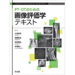
PT・OTのための画像評価学テキスト

出版社: 文光堂
著者:
発行日: 2022-06-17
分野: 臨床医学:一般  >  画像/超音波
ISBN: 9784830645983
電子書籍版: 2022-06-17 (第1版第1刷)
書籍・雑誌
≪全国送料無料でお届け≫
取寄せ目安:4~8営業日

5,280 円(税込)

電子書籍
章別単位での購入はできません
ブラウザ、アプリ閲覧

5,280 円(税込)

商品紹介

教育課程で使いやすい15章で構成され,運動器疾患・神経疾患・内部疾患をモダリティ別に解説.各分野のスペシャリストである医師と理学療法士により,解剖イラストと併せて疾患の特徴をよく反映したわかりやすい画像を数多く系統的に掲載.厳選された正常画像と疾患画像を対比しながら学習でき,読影のコツやピットホールの解説,学習の到達度を確認できるチャレンジ問題により評価能力を短期間で効率的に修得できるようになっている.

目次

  • Part 1 総論
     Chapter-1 基本画像診断・評価学

    Part 2 運動器疾患の画像
     Chapter-1 骨・関節X線の基本と画像解剖・評価
     Chapter-2 骨・関節CT・MRIの基本と画像解剖・評価
     Chapter-3 外傷の画像評価
     Chapter-4 関節疾患の画像評価
     Chapter-5 脊椎・脊髄疾患の画像評価
     Chapter-6 運動器エコーの基本と画像解剖・評価

    Part 3 神経疾患の画像
     Chapter-1 脳画像の基本と画像解剖・評価
     Chapter-2 脳血管疾患の画像評価1:出血
     Chapter-3 脳血管疾患の画像評価2:梗塞
     Chapter-4 脳血管疾患の画像評価3:その他

    Part 4 内部疾患の画像
     Chapter-1 胸部X線の基本と画像解剖・評価
     Chapter-2 胸部のCTの基本と画像解剖・評価
     Chapter-3 心血管のCT・MRIの基本と画像解剖・評価
     Chapter-4 心エコー・腹部エコーの基本と画像解剖・評価

この書籍の参考文献

参考文献のリンクは、リンク先の都合等により正しく表示されない場合がありますので、あらかじめご了承下さい。

本参考文献は電子書籍掲載内容を元にしております。

Part 2 運動器疾患の画像

P.45 掲載の参考文献
1) Magee, DJ : Orthopedic Physical Assessment, 5th ed, Saunders/Elsevier, Philadelphia, 58, 2008
2) 三木明徳 : 実習にも役立つ人体の構造と体表解剖, 金芳堂, 京都, 2016
3) 芳賀信彦 : 見過ごされがちな骨や関節の病気. 小児保健研究 70 : 598-602, 2011
4) 日本整形外科スポーツ医学会広報委員会監修 : スポーツ損傷シリーズ 4. 腰椎分離症. 三笠製薬, 2019
5) 三浦靖史ほか : 整形外科疾患の画像. 図解作業療法技術ガイド, 第4版, 石川 齊ほか編, 文光堂, 東京, 597-601, 2021
P.66 掲載の参考文献
1) 水間正澄ほか : 総論. リハビリテーション医療に活かす画像のみかた, 南江堂, 東京, 1-31, 2019
2) 山浦一郎 : 運動器リハビリテーションに必要な画像診断. 理学療法 40 : 310-313, 2013
3) Platzer W : 脊柱. 解剖学アトラス, 原著第10版, 平田幸男訳, 文光堂, 東京, 18-31, 2017
4) 松村讓兒ほか : 体幹の疾患. 病気がみえる vol. 11 運動器・整形外科, メディックメディア, 東京, 210-275, 2017
5) 寺山和雄ほか : 肩こり, 頸の痛み, 腕の痛み, 肩甲間部痛. 図で説く整形外科疾患, 医学書院, 東京, 1-14, 2005
6) Netter FH : 背部と脊髄等. ネッター解剖学アトラス, 原著第6版, 相磯貞和訳, エルゼビア・ジャパン, 東京, 152-177, 2016
7) 岡本嘉一ほか : スポーツ障害. 上肢の画像診断, メディカル・サイエンス・インターナショナル, 東京, 80-108, 2017
8) 内田淳正ほか : 手関節と手, 肩関節. 標準整形外科学, 第11版, 医学書院, 東京, 402-469, 2011
9) 川原康弘 : MRI による正常解剖. 股関節・骨盤の画像診断, メディカル・サイエンス・インターナショナル, 東京, 2-9, 2017
10) 新津守 : 膝の解剖. 膝MRI, 第3版, 医学書院, 東京, 1-13, 2018
11) 小橋由紋子 : 足関節・足部のMRI解剖. 足の画像診断, メディカル・サイエンス・インターナショナル, 東京, 3-14, 2013
P.85 掲載の参考文献
1) Heckman JD, et al : Acceleration of tibial fracture-healing by non-invasive, low-intensity pulsed ultrasound. J Bone Joint Surg Am 76 : 26-34, 1994
2) Neer CS 2nd : Four-segment classification of proximal humeral fractures : purpose and reliable use. J Shoulder Elbow Surg 11 : 389-400, 2002
3) Garden RS : Low-angle fixation in fractures of the femoral neck. J Bone Joint Surg Br 43B : 647-663, 1961
P.118 掲載の参考文献
1) 井村慎一ほか : 日本整形外科学会股関節機能判定基準. 日整会誌 69 : 860-867, 1995
4) 高岡邦夫ほか : 特発性大腿骨頭壊死症診断基準・病型・病期分類. 厚生労働省特定疾患対策事業骨関節系調査研究班, 平成12年度特発性大腿骨頭壊死症調査研究分科会報告書, 2001
P.143 掲載の参考文献
1) 遠藤健司ほか : 脊椎変形の画像診断. MB Orthopaedics 24 (増大号) : 110-116, 2011
2) 永島英樹 : 頸椎. 標準整形外科学, 第13版, 中村利孝ほか監, 井樋栄二ほか編, 医学書院, 東京, 499-527, 2017
3) 杜若陽祐 : 脊椎. 臨スポーツ医 22 : 65-72, 2005
4) 藤井正彦ほか : 脊椎疾患の画像診断 : MRIを中心にして. 断層映像研会誌 21 : 133-141, 1995
5) 永島英樹 : 胸椎, 腰椎. 標準整形外科学, 第13版, 中村利孝ほか監, 井樋栄二ほか編, 医学書院, 東京, 531-580, 2017
P.163 掲載の参考文献
1) Lee SH, et al : The usefulness of ultrasound and the posterior fat pad sign in pulled elbow. Injury 50 : 1227-1231, 2019
2) 工藤慎太郎 : 野球肘. 運動器疾患の「なぜ? 」がわかる臨床解剖学, 工藤慎太郎編, 医学書院, 東京, 53-63, 2012
3) Wiesler ER, et al : Ultrasound in the diagnosis of ulnar neuropathy at the cubital tunnel. J Hand Surg Am 31 : 1088-1093, 2006
4) 田中矢ほか : 上腕骨外側上顆炎における手関節背屈時の筋動態の変化 : 超音波検査装置を用いた滑走性評価. 日整外超音波会誌 28 : 114-119, 2016
5) Kiesel KB, et al : A comparison of select trunk muscle thickness change between subjects with low back pain classified in the treatment-based classification system and asymptomatic controls. J Orthop Sports Phys Ther 37 : 596-607, 2007
6) Takano Y, et al : Fat infiltration in the gluteus minimus muscle in older adults. Clin Interv Aging 13 : 1011-1017, 2018
7) Kara M, et al : Sonographic evaluation of sciatic nerves in patients with unilateral sciatica. Arch Phys Med Rehabil 93 : 1598-1602, 2012
8) Neto T, et al : Noninvasive measurement of sciatic nerve stiffness in patients with chronic low back related leg pain using shear wave elastography. J Ultrasound Med 38 : 157-164, 2019

Part 3 神経疾患の画像

P.194 掲載の参考文献
1) 寺島俊雄 : カラー図解 神経解剖学講義ノート, 金芳堂, 京都, 2011
2) Weinrich W, et al : 画像診断のための脳解剖と機能系, 久留裕ほか訳, 医学書院, 東京, 1995
3) Catani M, et al : Perisylvian language networks of the human brain. Ann Neurol 57 : 8-16, 2005
4) 石原健司 : 脳画像の読み方 : 基礎編. 高次脳機能研究 38 : 184-187, 2018
P.210 掲載の参考文献
1) Penfield W, et al : The Cerebral Cortex of Man, Macmillan, New York, 214-215, 1950
P.231 掲載の参考文献
1) Penfield W, et al : The Cerebral Cortex of Man, Macmillan, New York, 214-215, 1950
P.251 掲載の参考文献
1) 佐藤房郎 : 頭部外傷. ビジュアル実践リハ 脳・神経系リハビリテーション, 潮見泰藏編, 羊土社, 東京, 118-132, 2012
2) 渡邉修ほか : 脳外傷・低酸素脳症・脳腫瘍・水頭症. PT・OTビジュアルテキスト専門基礎 リハビリテーション医学, 安保雅博監, 渡邉修ほか編, 羊土社, 東京, 202-211, 2018
3) 渡邉修ほか : 脳出血. PT・OTビジュアルテキスト専門基礎 リハビリテーション医学, 安保雅博監, 渡邉修ほか編, 羊土社, 東京, 187-189, 2018
4) 藤井正純 : 脳腫瘍, 疾患別 : 評価と画像診断. MB Med Reha 223 : 104-118, 2018
5) Zucchella C, et al : Cognitive rehabilitation for early post-surgery inpatients affected by primary brain tumor : a randomized, controlled trial. J Neurooncol 114 : 93-100, 2013
6) Bartolo M, et al : Early rehabilitation after surgery improves functional outcome in inpatients with brain tumours. J Neurooncol 107 : 537-544, 2012
7) 望月久 : 多発性硬化症. ビジュアル実践リハ 脳・神経系リハビリテーション, 潮見泰藏編, 羊土社, 東京, 207-216, 2012

Part 4 内部疾患の画像

P.273 掲載の参考文献
1) 山口哲生 : 『人の肺』読影法とは. 見逃しなく読める! 胸部X線画像診断Q & A, 羊土社, 東京, 10-12, 2010
2) 松永尚文 : 心大血管・縦隔の異常. 新版 胸部単純X 線診断, 林邦昭ほか編, 秀潤社, 東京, 171-204, 2000
3) 佐々木成 : 浮腫と脱水, 濃縮と希釈の考え方. 日腎会誌 50 : 97-99, 2008
4) 本間光信 : 胸水貯留. リハビリテーションのための画像の読み方, 本間光信ほか編, メジカルビュー社, 東京, 126-128, 2015
5) 長尾大志 : 第3章 X線で黒くなる病態と白くなる病態. レジデントのためのやさしイイ胸部画像教室, 第2版, 日本医事新報社, 東京, 118-156, 2018
6) 山下敏彦 : 慢性閉塞性肺疾患. PT・OT のための画像のみかた, 山下敏彦監, 金原出版, 東京, 263-264, 2016
P.293 掲載の参考文献
1) 日本呼吸器学会COPDガイドライン第5版編集委員会 (編) : COPD (慢性閉塞性肺疾患) 診断と治療のためのガイドライン 2018, 第5版, 日本呼吸器学会, 東京, 2018
2) Cottin V, et al : Combined pulmonary fibrosis and emphysema : a distinct underrecognised entity. Eur Respir J 26 : 586-593, 2005
3) Raghu G, et al : Diagnosis of idiopathic pulmonary fibrosis. An Official ATS/ERS/JRS/ALATClinical Practice Guideline. Am J Respir Crit Care Med 198 : e44-e68, 2018
P.310 掲載の参考文献
1) Leipsic J, et al : SCCT guidelines for the interpretation and reporting of coronary CT angiography : a report of the Society of Cardiovascular Computed Tomography Guidelines Committee. J Cardiovasc Comput Tomogr 8 : 342-358, 2014
2) Jinzaki M, et al : Diagnostic accuracy of angiographic view image for the detection of coronary artery stenoses by 64-detector row CT : a pilot study comparison with conventional post-processing methods and axial images alone. Circ J 73 : 691-698, 2009
3) 安達秀雄 : 大動脈解離の分類. イラストレイテッド大動脈瘤手術, 金原出版, 東京, 60, 1999
4) Crawford ES, et al : Thoracoabdominal aortic aneurysms : preoperative and intraoperative factors determining immediate and long-term results of operations in 605 patients. J Vasc Surg 3 : 389-404, 1986
5) Takase K, et al : Demonstration of the artery of Adamkiewicz at multi- detector row helical CT. Radiology 223 : 39-45, 2002
P.333 掲載の参考文献
1) 日本循環器学会/日本心不全学会 : 急性・慢性心不全診療ガイドライン (2017年改訂版) https://www.j-circ.or.jp/cms/wp-content/uploads/2017/06/JCS2017_tsutsui_h.pdf (2021年12月閲覧)
2) 日本心エコー図学会 : ASE/EACVI ガイドラインおよび基準値 : 心エコーによる左室拡張機能評価のための勧告 : アメリカ心エコー図学会 (American Society of Echocardiography) およびヨーロッパ心血管画像学会 (European Association of Cardiovascular Imaging) による改訂 http://www.jse.gr.jp/contents/guideline/data/ASE-EACVIguideline&base.pdf
3) 日本循環器学会/日本胸部外科学会/日本血管外科学会/日本心臓血管外科学会 : 2020年改訂版弁膜症治療のガイドライン https://www.j-circ.or.jp/cms/wp-content/uploads/2020/04/JCS2020_Izumi_Eishi.pdf
4) 日本循環器学会/日本心不全学会 : 心筋症診療ガイドライン (2018年改訂版) https://www.j-circ.or.jp/cms/wp-content/uploads/2020/02/JCS2018_tsutsui_kitaoka.pdf (2021年12月閲覧)
5) 松尾汎 : 超音波による大動脈・末梢動脈病変の標準的評価法. 超音波医学 40 : 139-145, 2013
7) Terminology and Diagnostic Criteria Committee, Japan Society of Ultrasonics in Medicine : Standard method for ultrasound evaluation of aortic and peripheral arterial lesions. J Med Ultrason (2001) 41 : 535-546, 2014

最近チェックした商品履歴

Loading...